Triangles of Neck
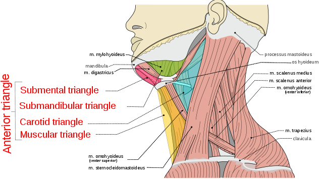
The following anatomic points define the anterior compartment/triangle of the neck:
- Superiorly: inferior border of the mandible
- Laterally: anterior border of the sternocleidomastoid muscle laterally
- Inferiorly: clavicle
- Medially: vertical midline from mental symphysis to suprasternal notch medially.
Contents: larynx, trachea, esophagus, thyroid and parathyroid glands, carotid sheath, and suprahyoid and infrahyoid strap muscles.
Submandibular triangle: region contained in the anterior neck bordered by the inferior margin of the mandible and the digastric, stylohyoid, and mylohyoid muscles. This region contains the submandibular gland and the marginal mandibular branch of the facial nerve.
Submental triangle: region bordered by the hyoid bone, the paired anterior bellies of the digastric muscles, and the mylohyoid muscle.
The upper belly of the omohyoid muscle in the anterior neck further divides the anterior neck into an upper carotid triangle and a lower muscular triangle.
The lateral neck or posterior triangle, is defined by following anatomic points:
- Medially: posterior aspects of the sternocleidomastoid muscle
- Laterally: trapezius muscle
- Inferiorly: middle 1/3 of clavicle
Contents: lymph node–bearing tissue, the spinal accessory nerve, and the cervical plexus.
The inferior belly of the omohyoid muscle further defines a lower subclavian triangle in the lateral neck that contains the brachial plexus and subclavian vessels.
General rules applicable to Neck Swellings
A. Rule of 7s for Duration of Swelling:
- If 7 days: Inflammatory
- If 7 months: Neoplastic
- If 7 years: Congenital
B. 80:20 rule for Malignant vs Benign Neck Swellings:
- In Pediatric age group: 20% are Malignant and 80% are Benign
- In Adult age group: 20% are Benign and 80% are Malignant
C. 20:40 rule for Age group:
<20 years:
- Congenital lesions: Thyroglossal cyst, Midline dermoid cyst, Branchial cyst, Cystic hygroma
- Inflammatory lymph nodes: Tonsillitis, Adenoids
- Chronic infections: Tuberculous lymph nodes
- Malignant lesions: Lymphoma, Metastatic lymph node (rarely)
20-40 years:
- Salivary gland pathology: Calculus, Infections, Tumors
- Thyroid pathology: Goiter, Neoplasms, Thyroiditis, Lymphoma
- Chronic infections: Tuberculosis, HIV
>40 years:
- Primary malignant tumor
- Metastatic lymph node
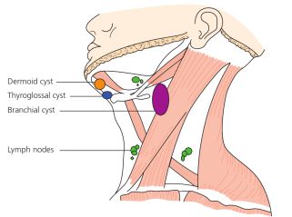
Midline Neck Swellings:
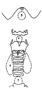
Submental region (2):
- Submental lymphadenitis
Between menton and hyoid (3):
- Ludwig’s angina
- Sublingual dermoid/Midline dermoid
- Ranula/Plunging ranula
- Thyroglossal cyst
Between hyoid and thyroid cartilage (4):
- Sub-hyoid bursitis
- Osteoma of hyoid bone
- Chondritis/Perichondritis
- Chondromas of thyroid cartilage
- Laryngocele
Between thyroid and cricoid (5):
- Delphian node enlargement
Between cricoid and supra-sternal notch (6):
- Thyroid gland – Goiters
At suprasternal space of Burns (7):
- Cold abscess/Lymph nodes
- Ectopic thyroid
- Suprasternal bursitis
- Neurofibroma of Supraclavicular nerve
- Aneurysm of arch of aorta
Laternal Neck Swellings:
| Anterior triangle | Posterior triangle | |
|---|---|---|
| Lymph nodes | Lymph nodes Cold abscess | Lymph nodes Cold abscess |
| Salivary glands | Submandibular swelling Parotid swelling | |
| Cystic structures | Branchial cyst | Cystic hygroma |
| Vascular structures | Carotid body tumor Carotid body aneurysm | Subclavian artery aneurysm |
| Other structures | Sternomastoid “tumor” | Cervical rib |
Mnemonic for Cystic neck swelling: ABCD
1. Anterior to SCM: Branchial cyst
2. Cystic hygroma: Dorsal to SCM
Solid and Cystic Swellings:
Solid swellings: Glands (Lymph node, salivary gland, thyroid gland), Vessels, Nerves, Subcutaneous, Sternocleidomastoid muscle, Bone
Cystic swellings: Air (Laryngocele), Fluid (Thyroglossal cyst, Dermoid cyst, Cystic hygroma, Branchial cyst), Abscess, Blood (Hemangioma, Aneurysm)
wow ! quite useful for revision and quick review 🙂
excellent presentation