Skip to content
Epomedicine

Mnemonics, Simplified Concepts & Thoughts

Epomedicine

Mnemonics, Simplified Concepts & Thoughts

Advanced glaucomatous changes diagram

Glaucomatous Optic Disc Changes Made Simple

Epomedicine, May 3, 2014May 5, 2014

Optic disc changes in Glaucoma is one of the most frequently asked topics in Ophthalmology. However, students are often found to have difficulty in understanding and remembering them. Hence, I came up with an idea to illustrate them with schematic diagrams and mnemonics to simplify the topic. The diagrams are conceptual only and may not coincide with the real images.

A. Pathophysiology of Optic Disc Changes in Primary Open Angle Glaucoma (POAG):

  1. Direct damage by pressure
  2. Capillary occlusion
  3. Interference with axoplasmic flow

B. Normal Optic Disc:
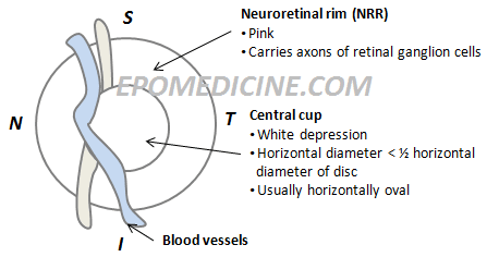
Normal optic disc drawing
Schematic drawing of normal optic disc

1. ISN’T rule:

  • Neuroretinal rim (NRR) is thickest inferiorly
  • NRR is thinnest temporally

2. Cup to Disc (C:D) ratio ~ 0.3 to 0.4

3. Asymmetry of C:D ratio in 2 eyes < 0.2

C. Early Glaucomatous Changes:
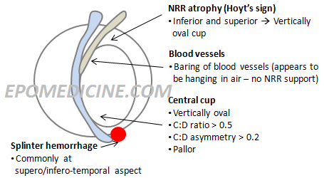
Early glaucomatous changes diagram
Schematic diagram of early optic disc changes in glaucoma

Mnemonic: VLAPS

  1. Vertically oval cup
  2. Large cup (C:D ratio > 0.6)
  3. Asymmetry of cups (Difference of C:D ratio > 0.2)
  4. Atrophy of NRR
  5. Pallor areas of disc
  6. Splinter hemorrhages

Note: ISN’T rule is no more applicable

D. Advanced Glaucomatous Changes:
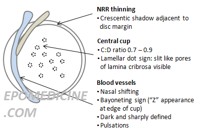
Advanced glaucomatous changes diagram
Schematic diagram of Advanced glaucomatous changes in optic disc

Mnemonic: B-LMNOP

  1. Bayonetting sign: double angulation of blood vessels
  2. Baring of circulinear blood bessels
  3. Laminar dot sign: Slit like pores of lamina cribrosa is visible
  4. Marked cupping: Cup to disc raio > 0.7 to 0.9
  5. Nasal shifting of blood vessels
  6. NRR thinning (Crescentic shadow adjacent to disc margin)
  7. Optic atrophy: This is the end stage where all NRR are destroyed, Optic Nerve Head (ONH) appears white and is deeply excavated
  8. Pulsation of retinal arterioles at disc margin

Submitted by:
Sulabh Shrestha
40 shares
  • Facebook40
  • Twitter
PGMEE, MRCS, USMLE, MBBS, MD/MS Ophthalmology

Post navigation

Previous post
Next post

Related Posts

PGMEE, MRCS, USMLE, MBBS, MD/MS

Acute Undifferentiated Febrile Illness – Mnemonic

Jun 8, 2021Jun 8, 2021

Acute undifferentiated febrile illnesses (AUFI) are characterised by fever of less than two weeks’ duration without organ-specific symptoms at the onset. Mnemonic MA-ESR lists the five main disease groups that cause acute undifferentiated febrile illnesses a. Malaria: Including all malaria due to Plasmodium falciparum, P vivax, P ovale, P malariae,…

Read More
PGMEE, MRCS, USMLE, MBBS, MD/MS respiratory center lesion

Respiratory Center Simplified

Jul 9, 2018

Mnemonic: Boat starts and PRASaD DIVEs 1. pre-Botzinger complex Starts respiratory rhythm (pacemaker): Boat Starts Location: Between nucleus ambiguus and lateral reticular nucleus (upper medulla) 2. Pneumotaxic center Represses Apneustic center: PR-A Location: Upper pons (Nucleus parabrachialis and Kolliker-fuse nucleus) 3. Apneustic center Stimulates DRG: A-SaD Location: Lower pons 4….

Read More
PGMEE, MRCS, USMLE, MBBS, MD/MS

Injection site swelling after vaccination in Infants

Mar 11, 2021Mar 11, 2021

Swelling and pain at injection site after vaccination is a frequently observed problem by parents and is often of concern to them. These type of reactions are generally observed after intramuscular vaccine and vaccine containing aluminium component. DPT as pentavalent is a frequent vaccine after which infants develop swelling. Types…

Read More

Comments (2)

  1. Syed S. Hasnain says:
    Jul 15, 2014 at 10:12 pm

    I hypothesize that glaucoma may not an optic disc neuropathy but an axotomy. As the disc/LC sinks the most peripheral fibers being closest to the scleral edge would be first to get stretched and severed followed by next in line central fibers. Severance of nerve fibers would lead to further sinking of the disc(due to loss of anchorage as roots anchor a tree)and thus severance of more of the nerve fibers. This cascade of sinking disc and severance of nerve fibers would continue until the central most fiber is severed in an orderly fashion. End-stage glaucoma disc may not be 100% cupped LC but a crater left over after the severance of all the nerve fibers.

    Reply

Leave a Reply Cancel reply

Your email address will not be published. Required fields are marked *

This site uses Akismet to reduce spam. Learn how your comment data is processed.

Epomedicine. Glaucomatous Optic Disc Changes Made Simple [Internet]. Epomedicine; 2014 May 3 [cited 2026 Jul 5]. Available from: https://epomedicine.com/medical-students/glaucomatous-optic-disc-changes-made-simple/.

Pre-clinical (Basic Sciences)

Anatomy

Biochemistry

Community medicine (PSM)

Embryology

Microbiology

Pathology

Pharmacology

Physiology

Clinical Sciences

Anesthesia

Dermatology

Emergency medicine

Forensic

Internal medicine

Gynecology & Obstetrics

Oncology

Ophthalmology

Orthopedics

Otorhinolaryngology (ENT)

Pediatrics

Psychiatry

Radiology

Surgery

RSS Ask Epomedicine

  • What to study for Clinical examination in Orthopedics?
  • What is the mechanism of AVNRT?

Epomedicine weekly

  • About Epomedicine
  • Contact Us
  • Author Guidelines
  • Submit Article
  • Editorial Board
  • USMLE
  • MRCS
  • Thesis
©2026 Epomedicine | WordPress Theme by SuperbThemes