Skip to content
Epomedicine

Mnemonics, Simplified Concepts & Thoughts

Epomedicine

Mnemonics, Simplified Concepts & Thoughts

Branches of Celiac trunk, Superior and Inferior mesenteric arteries and veins

Epomedicine, May 13, 2024May 13, 2024

Celiac trunk (T12)

Mnemonic: LHS (Left Hand Side)
Supplies Foregut (Upto opening of bile duct in 2nd part of duodenum)

1. Left gastric artery

  • Esophageal branches
  • Gastric branches

2. Hepatic artery (common)

  • Gastroduodenal artery
    • Supraduodenal artery
    • Superior pancreaticoduodenal artery
    • Right gastro-epiploic artery
  • Hepatic artery proper
    • Right gastric artery
    • Left hepatic artery
    • Right hepatic artery
      • Cystic artery

3. Splenic artery

  • Splenic branches
  • Pancreatic branches (body and tail)
  • Short gastric branches
  • Left gastro-epiploic artery

Superior Mesenteric Artery (L1)

Mnemonic: IMRIS
Supplies Midgut (Upto proximal 2/3 of transverse colon)

1. Inferior pancreaticoduodenal artery

2. Middle colic artery

3. Right colic artery

4. Ileo-colic artery

5. Small intestinal branches (Ileal and Jejunal)

Inferior Mesenteric Artery (L3)

Mnemonic: LeSS
Supplies Hindgut (Upto anal canal above pectinate line)

1. Left colic artery

2. Sigmoidal artery

3. Superior rectal artery

celiac trunk SMA IMA

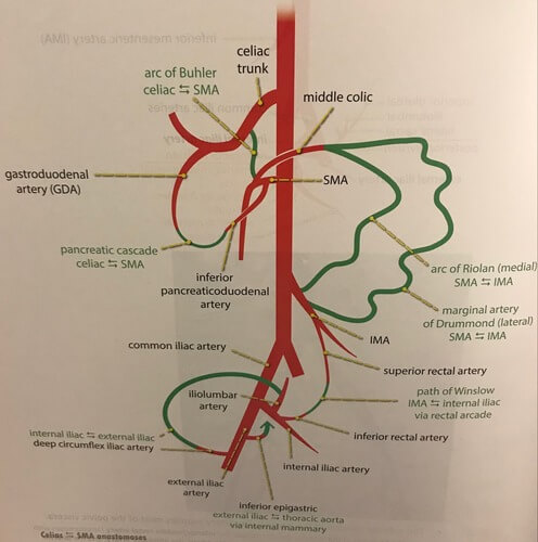
Collateral pathways:

The collateral pathways of the mesenteric arterial circulation include the pancreatico–duodenal arcade, arc of Riolan, arc of Buhler, arc of Barkow, and the marginal artery of Drummond.

  1. Kirk’s arcade: Dorsal pancreatic artery + Inferior pancreatico-duodenal arteries
  2. Arc of Buhler: Celiac artery + Superior mesenteric artery
  3. Arc of Barkow: Left gastroepiploic artery + Right gastroepiploic artery
  4. Arc of Riolan: Superior mesenteric artery (middle colic artery) + Inferior mesenteric artery
  5. Marginal artery of Drummond: Terminal branches of SMA + Terminal branches of IMA

Critical points:

  1. Sudeck’s critical point (rectosigmoid junction): At the point of origin of last sigmoidal arterial branch from IMA
  2. Griffith’s critical point (splenic flexure): At the site of watershed anastomosis between ascending left colic artery and marginal artery of Drummond

Venous counterparts

ArteriesVenous counterpartTributariesDrains into
Celiac arterySplenic veinShort gastric vein
Left gastro-epiploic vein
Pancreatic vein
Inferior mesenteric vein
Portal vein
Superior mesenteric arterySuperior mesenteric veinRight gastro-epiploic vein
Inferior pancreaticoduodenal veins
Jejunal vein
Ileal vein
Ileocolic vein
Right colic vein
Middle colic vein
Portal vein
Inferior mesenteric arteryInferior mesenteric veinSuperior rectal vein
Sigmoidal vein
Left colic vein
Splenic vein
Portal Vein : Tributaries and Portocaval Anastomoses
  • Facebook
  • Twitter
PGMEE, MRCS, USMLE, MBBS, MD/MS AnatomyCardiovascular systemGastrointestinal system

Post navigation

Previous post
Next post

Related Posts

PGMEE, MRCS, USMLE, MBBS, MD/MS implantation

Embryology Week 1: Cleavage to Implantation

Aug 2, 2016Aug 2, 2016

Following events occur in the zygote formed in the week zero of development: Cleavage Approximately 24 hours after fertilization the zygote begins with the first cleavage division. Series of mitotic divisions of the zygote (occurs in fallopian tube) to form small daughter cells called blastomeres. Characteristics of cleavage in humans:…

Read More
PGMEE, MRCS, USMLE, MBBS, MD/MS liver-segments-mnemonic

Liver Segments Explained with Mnemonic

Sep 8, 2016Jun 23, 2023

Couniaud divided liver into 8 functional segments, each of which is supplied by it’s own portal triad (composed of a portal vein, hepatic artery and a bile duct). Hepatic veins divide the liver in saggital plane: 1. Middle hepatic vein: Divides the liver into right and left functional lobe. 2….

Read More
PGMEE, MRCS, USMLE, MBBS, MD/MS

Intrinsic thumb muscles : Tricks to Remember

Apr 26, 2020Oct 27, 2022

The muscles of thumb will make 2 compartments – thenar compartment and adductor compartment. Mnemonic: Do it yourself as shown in the image below. Put your left hand with curled in middle/long finger above the palm of right hand. There are 2 muscle layers in this side of the hand….

Read More

Leave a Reply Cancel reply

Your email address will not be published. Required fields are marked *

This site uses Akismet to reduce spam. Learn how your comment data is processed.

Pre-clinical (Basic Sciences)

Anatomy

Biochemistry

Community medicine (PSM)

Embryology

Microbiology

Pathology

Pharmacology

Physiology

Clinical Sciences

Anesthesia

Dermatology

Emergency medicine

Forensic

Internal medicine

Gynecology & Obstetrics

Oncology

Ophthalmology

Orthopedics

Otorhinolaryngology (ENT)

Pediatrics

Psychiatry

Radiology

Surgery

RSS Ask Epomedicine

  • What to study for Clinical examination in Orthopedics?
  • What is the mechanism of AVNRT?

Epomedicine weekly

  • About Epomedicine
  • Contact Us
  • Author Guidelines
  • Submit Article
  • Editorial Board
  • USMLE
  • MRCS
  • Thesis
©2026 Epomedicine | WordPress Theme by SuperbThemes