A posterior hip dislocation puts the femoral head at risk of avascular necrosis (AVN) in between 5-60% of patients, depending on the delay in reduction and the severity of fracture-dislocation. The classic explanation is that the dislocated head kinks, stretches or tears the vessels that feed it.
A 2013 study from Warsaw (Zlotorowicz et al., Bone Joint J 2013;95-B:1453-7) used CT angiography after reduction – the authors mapped the arteries supplying the femoral head in 35 patients with posterior fracture-dislocation and compared them with 55 healthy hips.
Blood supply of femoral head in a normal hip
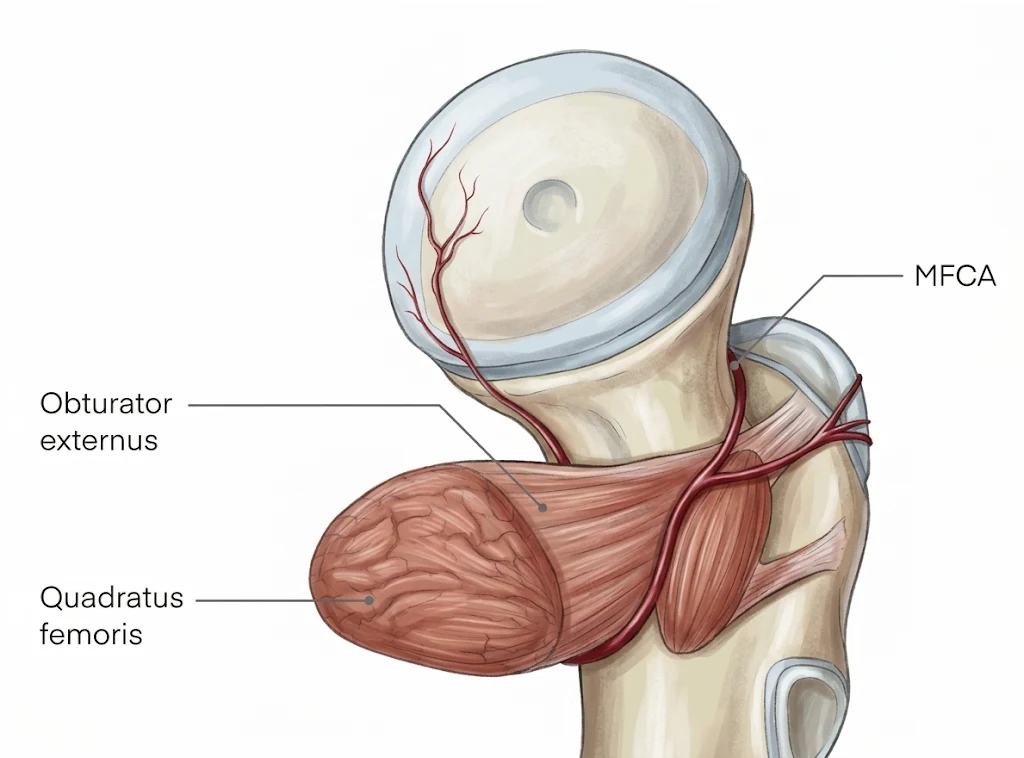
The major blood supply of femoral head comes from the medial femoral circumflex artery (MFCA)
- Deep branch of the MFCA: the dominant vessel. It ends as the postero-superior nutrient arteries, which enter the head through vascular foramina in the postero-superior and antero-superior quadrants of the head-neck junction.
- Postero-inferior nutrient artery: also arises from the MFCA, near its bifurcation, and runs towards the superior margin of the lesser trochanter.
- Piriformis branch of the inferior gluteal artery: reaches the deep branch of the MFCA through an anastomosis.
Two minor contributors are the anterior nutrient artery of the femoral neck (from the lateral femoral circumflex artery) and the artery of the ligamentum teres (from the obturator artery).
Why posterior dislocation threatens the head
The deep branch of the MFCA runs posterior to obturator externus and anterior to quadratus femoris, then pierces the capsule at the posterior trochanteric region. In a posterior dislocation the displaced head can kink or stretch it at exactly this point.
The Kocher-Langenbeck approach used to fix the acetabular fracture works in the same posterior territory. That makes the piriformis branch of the inferior gluteal artery vulnerable to the injury and to the surgery.
The 2013 Warsaw study
The authors analysed CT angiograms from 35 hips (34 men, 1 woman; mean age 37.1 years, range 16-64) with unilateral posterior fracture-dislocation (20 left, 15 right). All scans were done after reduction, as part of pre-operative planning. The control group was 55 healthy hips from their earlier study.
- Fracture pattern: posterior wall plus dislocation in 57%, transverse plus posterior wall in 37%, posterior column in 6%.
- Time to reduction: under 12 hours in 49%, 12 to 24 hours in 14%, over 24 hours in 37%.
- Treatment: closed reduction followed by ORIF through a Kocher-Langenbeck approach in 33 patients (94%); closed reduction alone in 2.
- Imaging: 64-section CT, 0.6 mm slices, 90-120 ml of iodinated contrast as a rapid bolus, scanned 25-30 seconds after injection to catch the middle-to-late arterial phase. Images were read with axial scans, multiplanar reformats, volume rendering and maximum intensity projection.
- Outcome: 10 of the 35 patients developed AVN at a minimum follow-up of 12 months (diagnosed on average 5.2 months after injury, range 1-9).
Key findings
The deep branch of the MFCA was still carrying contrast after reduction in 91% of injured hips, and it was larger than in healthy hips. The piriformis branch was the vessel that was lost most often.
| Finding | Fracture-dislocation (n = 35) | Control (n = 55) | p |
|---|---|---|---|
| Deep branch of MFCA filled with contrast | 32 (91%) | 53 (96%) | 0.37 |
| Deep branch diameter, 10 mm from bifurcation (mean, range) | 1.8 mm (1.1-2.7) | 1.6 mm (1.1-2.7) | 0.022 |
| Postero-inferior nutrient artery filled | 8 (23%) | 6 (11%) | 0.13 |
| Piriformis branch of inferior gluteal artery filled | 9 (26%) | 27 (49%) | 0.027 |
Deep branch. Flow was present in 32 hips, which is not significantly different from controls. Where it was absent, CT angiography showed where contrast stopped. In one patient it stopped near the MFCA bifurcation, and in two others at 26 mm and 35 mm along the deep branch. In one hip a fractured bone fragment near the inferior femoral neck blocked the main MFCA trunk, yet the deep branch still filled through an anastomosis with the obturator artery via the superficial branch of the MFCA.
Larger calibre. The deep branch was significantly wider in injured hips (p = 0.022). The authors read this as increased flow during revascularisation after an episode of ischaemia. The terminal postero-superior nutrient arteries were too small to see.
Piriformis branch. It was seen in only 26% of injured hips against 49% of controls (p = 0.027). The authors suggest that the dislocation and the Kocher-Langenbeck exposure can both injure this vessel. The postero-inferior nutrient artery was too small to measure, and its difference from controls was not significant.
The AVN subgroup
Ten patients developed AVN, and every one of them had waited more than 12 hours for reduction (three a mean of 13 hours, seven a mean of 15 days). Their angiograms were not what most clinicians would predict.
- Deep branch: contrast-enhanced in 8 of 10 hips (80%), with a mean diameter of 1.8 mm (1.4-2.4), again larger than in controls.
- Postero-inferior nutrient artery: seen in 4 hips (40%).
- Inferior gluteal anastomosis: seen in 1 hip.
- No flow in any of the three main vessels: 2 patients, both of whom developed AVN. Their reductions were delayed by 13 and 40 days, and flow was never restored.
Of the three patients with no flow in the deep branch, two developed AVN (the two above) and one did not. The patient who escaped had a large postero-inferior nutrient artery, which may have helped preserve the head.
The authors did no statistical testing in this subgroup because the numbers were too small.
What does it mean?
Reduction restores flow in most hips. Deep branch of MFCA was patent in 91%. Once the hip is reduced, tension on the vessel is relieved and perfusion returns. This is consistent with the importance of prompt reduction, although the study did not test timing directly.
A bigger vessel may mean a past ischaemic insult. The wider deep branch in injured hips, including those with AVN, may reflect revascularisation after ischaemia, or even sub-clinical AVN with increased flow. The authors point out that this matches the increased uptake seen on scintigraphy in the later phase of AVN. They also note that all ten AVN patients waited more than 12 hours, so initial ischaemia while the hip was dislocated is a plausible cause.
Patent vessels do not exclude AVN. Eight of the ten patients who developed AVN had an enhancing deep branch at the time of the scan. Flow after reduction tells you the vessel is open, not that the head is healthy.
Absent flow on all three vessels is a bad sign. Both patients with no enhancement in any of the three main arteries developed AVN. The authors suggest CT angiography could help predict the prognosis of the hip after posterior dislocation, and it can show the level at which the vessel is blocked.
The posterior approach has a cost. The reduced piriformis branch filling in injured hips fits with the idea that the injury and the Kocher-Langenbeck exposure can both damage this contribution.
Limitations
- The scans were taken after reduction, so we do not know what the blood flow was while the hip was dislocated.
- Controls were a historical group of healthy hips, not the uninjured contralateral side of the same patients.
- Only 10 patients had AVN, and 2 had absent flow in all vessels, so the AVN findings are descriptive and untested statistically.
- Vessel diameter is an indirect marker of flow, and the smallest vessels (the postero-superior terminal branches and the postero-inferior nutrient artery) could not be measured.
- There was one woman, and the cohort is mostly young men from high-energy trauma, which limits generalisability.
- Contrast CT angiography adds contrast and radiation exposure, which are worth weighing before using it routinely.
Take-home points
- CT angiography after reduction can show the three main arteries of the femoral head, and where contrast stops when a vessel is blocked.
- The deep branch of the MFCA was open in 91% of hips after reduction of a posterior fracture-dislocation, and it was wider than in healthy hips (1.8 vs 1.6 mm, p = 0.022).
- The piriformis branch of the inferior gluteal artery was seen less often in injured hips (26% vs 49%, p = 0.027).
- All 10 patients who developed AVN had a delay to reduction of more than 12 hours.
- Both patients with no flow in any of the three main vessels developed AVN. Eight of the ten AVN patients still had an open deep branch.
- The study is small and descriptive. It cannot say what happened to blood flow while the hip was dislocated.
Reference
Zlotorowicz M, Czubak J, Caban A, Kozinski P, Boguslawska-Walecka R. The blood supply to the femoral head after posterior fracture/dislocation of the hip, assessed by CT angiography. Bone Joint J 2013;95-B(11):1453-1457. doi:10.1302/0301-620X.95B11.32383

He is the section editor of Orthopedics in Epomedicine. He searches for and share simpler ways to make complicated medical topics simple. He also loves writing poetry, listening and playing music. He is currently pursuing Fellowship in Hip, Pelvi-acetabulum and Arthroplasty at B&B Hospital.