Auditory Pathway Component Mnemonic
E.C.O.L.I.M.A
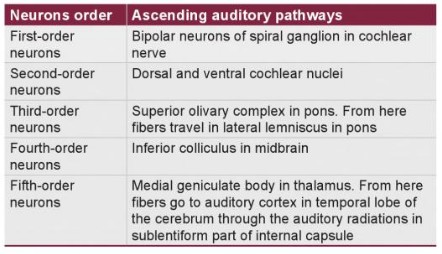
Ascending from peripheral to central the components are:
- Ear receptors (Hair cells) in Cochlea and Eighth Cranial nerve (CN VIII)
- Cochlear nucleus
- Superior Olivary nucleus
- Lateral lemniscus
- Inferior colliculus
- Medial geniculate body
- Auditory cortex
Explanation of the Mnemonic
Ear receptors and Eighth cranial nerve (Organ of Corti)
- Cochlear hair cells are located on basilar membrane (In Organ of Corti):
- At the base of cochlea (thinner basilar membrane): encodes high-frequency sounds
- At the apex of cochlea (thicker basilar membrane): encodes low-frequency sounds
Remember a guitar and compare the strings with the basilar membrane – Thinner strings are more tight and produce high frequency like in base of cochlea and Thicker strings are loose and produce low frequency sounds like in apex of cochlea. This localization of sound by cochlea is known as Tonotopic localization.
- Sound waves entering scala vestibuli and back to scala tympani causes vibration of basilar membrane.
- Vibration of basilar membrane leads to movement of sterocilia of hair cells against the tectorial membrane that leads to generation of action potential and production of electrical impulses that travel down the CN VIII (cochlear division).
- Axons of spiral ganglion bipolar axons form the cochlear nerve.
Cochlear Nuclei (Pontomedullary junction)
- Cochlear nuclei is divided into 2 divisions: Ventral cochlear nuclei and Dorsal cochelar nuclei in relation to the inferior cerebellar peduncle in Ponto-medullary junction.
- Anterior (ventral) cochlear nucleus is dedicated to low frequency (Apical) sounds.
- Posterior (dorsal) cochlear nucleus is dedicated to high frequency (Basal) sounds.
- Anterior cochlear nucleus → Anterior acoustic stria → Bilateral superior olivary nucleus (Binaural sound localization pathway)
- Crossing over of anterior acoustic stria due to bilateral projection form a trapezoidal shape (trapezoid body).
- Posterior cochlear nucleus → Posterior acoustic stria → Contralateral lateral lemniscus and inferior colliculus (Monoaural sound localization pathway)
- Skips superior olivary nucleus
Superior Olivary Nuclei (Pons)
- Located in pons
- 1st auditory nuclei to reveive binaural input and use the binaural input to localize sound forces.
Note: Inferior olivary nucleus is involved in motor control and provides major projection to the cerebellum.
Lateral Lemniscus (Midbrain)
- Projection fibers from Ipsilateral Superior olivary nucleus.
Note: Medial lemniscus is a part of dorsal column pathway.
Inferior Colliculus (Midbrain)
- Receives fibers from ipsilateral superior olivary nuclei through lateral lemniscus
Note: Superior colliculus is a part of visual pathway.
Medial Geniculate Body (Thalamus)
- Receives projections from ipsilateral inferior colliculus via brachium of inferior colliculus
Note: Lateral Geniculate body is a part of visual pathway.
Read: Mnemonic for Thalamic connections
Auditory Cortex
- Auditory radiations from MGB to respective transverse gyri of Heschl (Brodmann areas 41 and 42), deep to the superior
temporal gyrus in the Sylvian fissure via sublentiform part of internal capsule. - The auditory association cortex surrounds the primary auditory cortex.
Clinical Relevance
Since, the projection is bilateral to the superior olivary nucleus –
- Lesions of cochlear part of CN VIII or cochlear nuclei at pontomedullary junction cause profound unilateral sensory hearing loss.
- All other lesions to the auditory pathway above the cochlear nuclei cause bilateral suppression of hearing and decreased ability to localize a sound source.

He is the section editor of Orthopedics in Epomedicine. He searches for and share simpler ways to make complicated medical topics simple. He also loves writing poetry, listening and playing music. He is currently pursuing Fellowship in Hip, Pelvi-acetabulum and Arthroplasty at B&B Hospital.