Spaces of Hand
Deep space infections occur in one of the three anatomically defined potential spaces within the hand.
- Thenar, mid-palmar and hypothenar spaces
- Interdigital subfascial web space
- Forearm space of parona
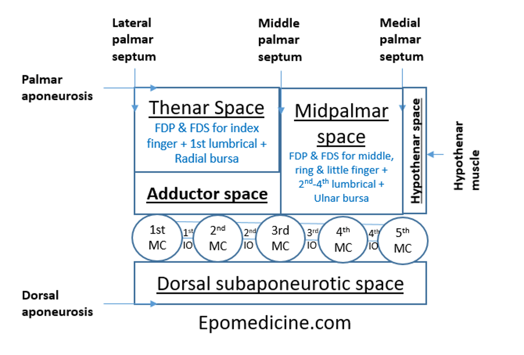
| Deep hand space | Dorsal border | Volar border | Ulnar border | Radial border | Remarks |
| Thenar | Adductor pollicis | Palmar aponeurosis | Middle palmar septum | Lateral palmar septum | FPL within synovial sheath, FDP and FDS for index finger, 1st lumbrical, Radial bursa, Palmar digital vessels of thumb and lateral side of index finger – these can be considered as contents or volar border |
| Mid-palmar | 3rd, 4th and 5th metacarpals with fascia covering 3rd and 4th interossei | Palmar aponeurosis | Medial palmar septum | Middle palmar septum | Flexor tendons of 3rd-5th fingers, 2nd-4th lumbricals, Superficial palmar arch, Digital nerves and vessels of medial 3 and 1/2 fingers, Ulnar bursa – these can be considered as contents or volar border |
| Hypothenar | 5th metacarpal | Palmar aponeurosis | Medial palmar septum | Hypothenar muscles | |
| Dorsal subaponeurotic | Extensor tendons and dorsal aponeurosis | Metacarpals and dorsal fascia of interossei | |||
| Interdigital subfascial | Dorsal hand fascia and skin | Palmar fascia | Metacarpophalangeal joint and extensor tendon | Metacarpophalangeal joint and extensor tendon | |
| Parona (Forearm space) | Digital flexor tendons | Pronator quadratus | Flexor carpi ulnaris | Flexor pollicis longus | Continuous with midpalmar space |
Incisions:
- Mid-palmar space: Curved incision beginning at the level of distal palmar crease, in line with the long finger and extending ulnar-ward to just inside the hypothenar eminence
- Thenar space: Curved incision in thumb web parallel to border of the 1st dorsal interossei or along the medial side of thenar crease

Compartments of Hand
Hand comprises of 10 compartments.
| Compartment | Muscles | Fasciotomy Incision |
|---|---|---|
| Hypothenar | Abductor digiti minimi Flexor digiti minimi Opponens digiti minimi | Ulnar border of hand |
| Thenar | Abductor pollicis brevis Oponens pollicis Flexor pollicis brevis | Radial margin of thenar eminence |
| Adductor | Two heads of adductor pollicis | Dorsal over first webspace |
| Dorsal Interosseous | 4 compartments | Between 2-3rd & 4-5th metacarpals |
| Volar Interosseous | 3 compartments | Extended carpal tunnel incision |
3 Dorsal fasciotomy incisions:
Incision 1: Longitudinal Incision in 1st Webspace
- First dorsal interosseous muscle (can also release the dorsal fascia of the adductor pollicis)
Incision 2 & 3: Longitudinal Incision in 2nd-3rd, 4-5th Metacarpal space
- Remaining dorsal interosseous muscles (can also decompression volar interosseus muscles)