Skip to content
Epomedicine

Mnemonics, Simplified Concepts & Thoughts

Epomedicine

Mnemonics, Simplified Concepts & Thoughts

Anatomy and Physiology of Sebaceous glands

Epomedicine, Nov 11, 2013

Definition: Sebaceous glands are numerous microscopic glands in the dermis that usually open into the hair follicles and secrete sebum. They are holocrine glands, i.e., the sebum consists of the entire secreting cells.

Location: Found everywhere on the skin apart from the palms and soles

Types of Sebaceous glands:

1. Associated with hair follicles in hair covered areas:

  • Numerous and prominent on the head, neck, chest and back
  • These lie in the obtuse angle between the follicle and the epidermis
  • Discharge their secretions into the ducts of hair follicles

2. Free/Modified sebaceous glands independent of hair follicles:

  • Seen at sites devoid of hair (glaborous skin) – mucosal margins
  • These open directly on the surface
  • Found in:
    • Eyelid (meibomian glands)
    • Margins of lip (Fordyce spot)
    • Areola of nipples (Montgomery tubercles)
    • Prepuce of penis (Tyson glands)
    • Mucocutaneous surface of female genitalia
    • Peri-anal region

Histology of Sebaceous gland:

Sebaceous gland anatomy

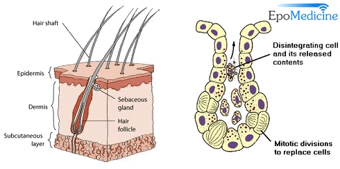
1. Multilobed gland: made up of lipid containing cells, which secrete sebum as a holocrine secretion

2. Duct: usually opens into hair follicles

Composition of sebum: triglycerides, fatty acids, wax esters, squalene and cholesterol

Note: Free fatty acids are produced by breakdown of triglycerides by lipases secreted by bacteria – Propionibacterium acnes

Functions of sebum:

  1. Lubricant for skin
  2. Mild bacteriocidal and fungistatic activity
  3. Vitamin D precursor and Vitamin E production

Variation in sebum secretion:

  • Sebum secretion varies between body regions and individual to individual.
  • Sebum output in descending order: scalp and forehead, face, back and chest, axilla, pubis, neck, abdomen, lower arm, leg, back of hand and ankle

Hormonal control of Sebaceous glands:

Sebaceous hormonal control

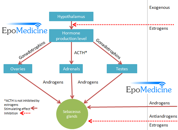
Androgenic hormones, especially dihydrotestosterone, stimulate sebaceous gland activity but it is not the level of circulating androgens which is important, but an enhanced end organ sensitivity. Human sebaceous glands contain 5α reductase, 3α- and 17α-hydroxysteroid dehydrogenase, which convert weaker androgens to dihydrotestosterone, which in turn binds to specific receptors (PPARs and Melanocortin receptors) in sebaceous glands, increasing sebum secretion.

At birth the sebaceous glands are functional, but after that they become less active until puberty.

Growth hormone and thyroid hormone also affects sebum production.

Disorders of Sebaceous gland:

  1. Acne
  2. Rosacea
11 shares
  • Facebook11
  • Twitter
PGMEE, MRCS, USMLE, MBBS, MD/MS DermatologyMusculoskeletal system

Post navigation

Previous post
Next post

Related Posts

PGMEE, MRCS, USMLE, MBBS, MD/MS thalamus structure

Thalamic Connections Mnemonic

Jul 16, 2016Aug 4, 2023

Structure of Thalamus A vertical “Y” shaped white mater – internal medullary lamina divides thalamus into: In anatomical position: Pulvinar = Posterior end or posterior pole of thalamus Thalamic Connections Picture mnemonic Remember the schematic diagram drawn below showing important parts of thalamus in an anticlockwise fashion: Now, we assign…

Read More
PGMEE, MRCS, USMLE, MBBS, MD/MS

Popliteal Fossa : Mnemonics

May 13, 2023Aug 9, 2023

Mnemonic: Serve And Volley Next Ball Think of popliteal fossa as a diamond-shaped space. The floor is formed by Popliteus muscle. Membranosus = Medial (The 2 “Semis” go together – hence medial border is formed by semimembranosus, semitendinosus and medial head of gastrocnemius) Therefore, biceps femoris and lateral head of…

Read More
PGMEE, MRCS, USMLE, MBBS, MD/MS hilum signs

Chest Xray – Approach to hilum

Jun 24, 2015Jun 24, 2015

Hilum in human anatomy refers to the depression where structures such as blood vessels and nerves enter an organ. The structures contributing to hilar shadows in a Chest X-ray are: Major: Pulmonary artery and veins Minor: Fat, Lymph nodes and Bronchial walls Normal Hilum: Position: Left hilum is slightly higher…

Read More

Leave a Reply Cancel reply

Your email address will not be published. Required fields are marked *

This site uses Akismet to reduce spam. Learn how your comment data is processed.

Pre-clinical (Basic Sciences)

Anatomy

Biochemistry

Community medicine (PSM)

Embryology

Microbiology

Pathology

Pharmacology

Physiology

Clinical Sciences

Anesthesia

Dermatology

Emergency medicine

Forensic

Internal medicine

Gynecology & Obstetrics

Oncology

Ophthalmology

Orthopedics

Otorhinolaryngology (ENT)

Pediatrics

Psychiatry

Radiology

Surgery

RSS Ask Epomedicine

  • What to study for Clinical examination in Orthopedics?
  • What is the mechanism of AVNRT?

Epomedicine weekly

  • About Epomedicine
  • Contact Us
  • Author Guidelines
  • Submit Article
  • Editorial Board
  • USMLE
  • MRCS
  • Thesis
©2026 Epomedicine | WordPress Theme by SuperbThemes