Skip to content
Epomedicine

Mnemonics, Simplified Concepts & Thoughts

Epomedicine

Mnemonics, Simplified Concepts & Thoughts

Schirmer’s test

Epomedicine, Jun 26, 2020Jun 26, 2020

Schirmer’s test is a measurement of tear production, devised by the German ophthalmologist Otto W.A. Schirmer. Originally, Schirmer used blotting paper to collect tears elicited by one of the 3 methods of stimulating lacrimation:

  1. Inserting the strip of paper itself
  2. Irritating the nasal mucosa after the cornea had been anesthetized
  3. Having the patient look into the sun

Indication: Evaluation of dry eye (Measures aqueous tear production)

Mnemonic:

Test 1 measures 2 things.
Test 2 measures 1 thing.
Test 3 measures Nose thing.

Types:

schirmer test

1. Schirmer I (without anesthetic): measures baseline and reflex tear secretion

  • function of main lacrimal gland, whose secretory activity is stimulated by the irritating nature of filter paper

2. Schirmer II (with anesthetic): measures baseline secretion

  • function of accesory lacrimal glands (the basic secretors)

3. Schirmer III (without anesthetic): with nasal irritation

  • nasal mucosal stimulation represents a maximal secretory stimulation for the lacrimal glands and can thus reveal the maximal secretory capacity of the lacrimal glands.

According to some authors: Schirmer I is divided into a. without anesthetic and b. with anesthetic. While Schirmer II represents test with nasal irritation.

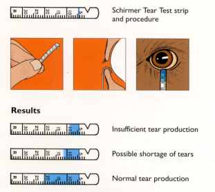
Materials required: 5 X 35 mm of Schirmer’s strip or Whatman filter paper no. 41

Procedure:

  1. The eye is gently dried of excess tears
  2. The schirmer strip is folded 5 mm from one end and kept in the lower fornix at the junction of lateral 1/3 and medial 2/3 (do not touch cornea or lashes)
  3. The patient is asked to close the eyes.
  4. Tears in the conjunctival sac will cause progressive wetting of the paper strip.
  5. After 5 minutes, the filter paper is removed and the distance between the leading edge of wetness and the initial fold is measured, using a millimeter ruler.

Interpretation:

Normal: >15 mm

Mild-moderate Keratoconjunctivitis sicca (KCS): 5-10 mm

Severe KCS: <5 mm

<10 mm is considered abnormal in Schirmer 1 and <5 mm in Schirmer 2

Causes of KCS:

  1. Idiopathic
  2. Congenial alacimia
  3. Xerophthalmia
  4. Lacrimal gland ablation
  5. Sensory denervation
  6. Collagen vascular disease: Sjogren syndrome, SLE, RA
5 shares
  • Facebook5
  • Twitter
Clinical Skills and Approaches Clinical examinationOphthalmology

Post navigation

Previous post
Next post

Related Posts

Clinical Skills and Approaches Bryant's triangle

Bryant’s triangle (Examination of Hip)

Jun 4, 2014Sep 5, 2022

Synonyms: Iliofemoral triangle Position: The patient lies supine with the pelvis square, and the limbs in identical position. Points of the Bryant’s triangle: The Bryant’s triangle is a right angled formed by: Anterior Superior Iliac Spine (ASIS) Tip of Greater trochanter Junction of perpendiculars from the 2 points above (1st draw…

Read More
Clinical Skills and Approaches history and examination format

Cyanosis

Dec 3, 2013Feb 16, 2018

Synonyms: Morbus coeruleus, Blue disease Definition of Cyanosis Cyanosis is the bluish discoloration of the skin, mucous membrane and nail bed usually owing to atleast 5 gm/dl of reduced hemoglobin/deoxyhemoglobin or abnormal hemoglobin derivatives (eg. methemoglobin, sulfhemoglobin, etc.) circulating within the superficial dermal capillaries and subpapillary venous plexus (not, as commonly taught, the…

Read More
Clinical Skills and Approaches ascitic paracentesis site

Ascitic Paracentesis : Practical Essentials

Jan 16, 2017

Absolute contraindications to Ascitic Tapping (Paracentesis) While some authors have claimed ascitic paracentesis to be free from absolute contraindications, others have listed following as absolute contraindications: Clinically evident fibrinolysis Disseminated Intravascular Coagulation (DIC) Clinically apparent oozing from needle sticks Acute abdomen requiring surgery Relative contraindications to Blind Paracentesis Ultrasound guided…

Read More

Comment

  1. Girma says:
    Dec 8, 2020 at 6:09 am

    The above test types have some disagreements with what is written on “2019-2020 BCSC 08 External Disease and Cornea” books

    Reply

Leave a Reply Cancel reply

Your email address will not be published. Required fields are marked *

This site uses Akismet to reduce spam. Learn how your comment data is processed.

Pre-clinical (Basic Sciences)

Anatomy

Biochemistry

Community medicine (PSM)

Embryology

Microbiology

Pathology

Pharmacology

Physiology

Clinical Sciences

Anesthesia

Dermatology

Emergency medicine

Forensic

Internal medicine

Gynecology & Obstetrics

Oncology

Ophthalmology

Orthopedics

Otorhinolaryngology (ENT)

Pediatrics

Psychiatry

Radiology

Surgery

RSS Ask Epomedicine

  • What to study for Clinical examination in Orthopedics?
  • What is the mechanism of AVNRT?

Epomedicine weekly

  • About Epomedicine
  • Contact Us
  • Author Guidelines
  • Submit Article
  • Editorial Board
  • USMLE
  • MRCS
  • Thesis
©2026 Epomedicine | WordPress Theme by SuperbThemes