Skip to content
Epomedicine

Mnemonics, Simplified Concepts & Thoughts

Epomedicine

Mnemonics, Simplified Concepts & Thoughts

spleen percussion

Percussion of Spleen

Epomedicine, Jan 31, 2017

Traube’s semilunar space

Borders:

  1. Superiorly: Left 6th rib superiorly
  2. Laterally: Left midaxillary line or Left anterior axillary line
  3. Inferiorly: Left costal margin

Method:

  1. Patient’s position: supine with left arm slightly abducted.
  2. Percuss: from medial to lateral

Interpretation: Resonance (Normal) and Dullness (Splenomegaly)

  • Also: Pleural effusion or mass in stomach may cause dullness in Traube’s space.

Castell’s method

Patient’s position: Supine

Percuss: In the lowest intercostal space in the anterior axillary line (8th or 9th)

Interpretation:

  1. Resonant percussion note on expiration or full inspiration: normal
  2. Dull percussion note on full inspiration: splenomegaly
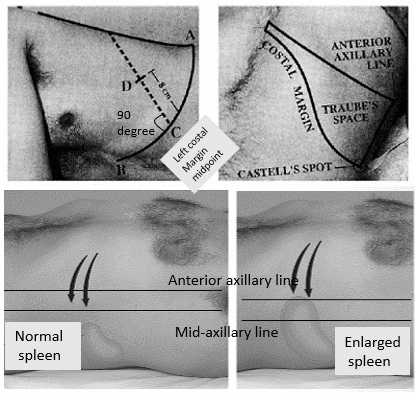
spleen percussion
Top-left: Nixon’s method; Top-right: Traube’s space and Castell’s method; Bottom: Line of splenic enlargement

Nixon’s method

Patient’s position: Right lateral decubitus (rationale: spleen comes to lie above colon and stomach).

Percuss: start percussing midway along the left costal margin and proceed in a line perpendicular to left constal margin

Interpretation: Upper limit of dullness >8 cm above costal margin: Splenic enlargement

25 shares
  • Facebook23
  • Twitter
Clinical Skills and Approaches Clinical examinationGastrointestinal systemInternal medicine

Post navigation

Previous post
Next post

Related Posts

Clinical Skills and Approaches

Splenomegaly : Examination techniques and Clinical Approach

Mar 20, 2021May 31, 2023

ANATOMY OF SPLEEN Histologically: FUNCTIONS OF SPLEEN Spleen is the largest lymphoid organ organ and serves following functions – An increase in these normal functions may result in splenomegaly EXAMINATION OF SPLEEN 1. Palpation: If history suggest splenomegaly but is not palpable: Roll the patient on to the right lateral…

Read More
Clinical Skills and Approaches opioid algorithm

Prescribing Opioids for Chronic Pain

Jan 12, 2017Jan 12, 2017

Recommendations for Prescribing Opioid in Chronic pain 1. 1st line of therapy in chronic pain outside of active cancer, palliative or end of life care must be nonpharmacologic therapy and nonopioid pharmacologic. Consider opioid therapy in combination with nonpharmacologic or nonopioid pharmacologic therapy if benefits outweighs risk. 2. Ordinarily 2…

Read More
Clinical Skills and Approaches

WIPERS mnemonic for starting patient examination

Jun 20, 2021Oct 16, 2022

A useful approach before examining any patient, using the mnemonic WIPER. 1. Wind up clothes above elbow, Withdraw all jewelry and Wash your hands. 2. Introduce yourself. 3. Purpose and Permission – explain purpose and gain verbal consent (permission) to proceed with the examination. 4. Expose the patient as needed….

Read More

Comments (3)

  1. nagabushana m.v. says:
    Dec 18, 2022 at 4:53 pm

    As for Nixon’s method- if dullness is beyond 8cms on a perpendicular line from middle of left costal margin,it means spleen is enlarged upto costal margin.This can be easily picked up by hooking method or Medleton’s manuare.Nixon’s method becomes superfluous.
    Well ,dullness lower anterior chestwall may indicate pleural effusion or pleural thickening or even a fundal growth in the stomach .We need to ponderon that.

    Reply
  2. Purusottam Misra says:
    Mar 17, 2023 at 3:00 pm

    A simple ultrasound
    Willsufice for 100 percent diagnosis

    Reply
    1. Epomedicine says:
      Mar 17, 2023 at 4:21 pm

      1. Physical examination for splenomegaly have sensitivity ranging from 11 to 85 % and specificity from 32 to 99 %. Physical examination plus POCUS had a sensitivity of 100 % and specificity of 74 %.

      Reference: Olson AP, Trappey B, Wagner M, Newman M, Nixon LJ, Schnobrich D. Point-of-care ultrasonography improves the diagnosis of splenomegaly in hospitalized patients. Crit Ultrasound J. 2015 Dec;7(1):13. doi: 10.1186/s13089-015-0030-8. Epub 2015 Sep 17. PMID: 26383010; PMCID: PMC4574040.

      2. Sonographic versions of traditional physical examination maneuvers have greater diagnostic accuracy than the physical examination maneuvers from which they are derived but may take longer to perform. We recommend a combination of traditional physical examination and sonographic techniques when evaluating for splenomegaly at the bedside.

      Reference: Cessford T, Meneilly GS, Arishenkoff S, Eddy C, Chen LYC, Kim DJ, Ma IWY. Comparing Physical Examination With Sonographic Versions of the Same Examination Techniques for Splenomegaly. J Ultrasound Med. 2018 Jul;37(7):1621-1629. doi: 10.1002/jum.14506. Epub 2017 Dec 8. PMID: 29219201.

      Reply

Leave a Reply to Purusottam Misra Cancel reply

Your email address will not be published. Required fields are marked *

This site uses Akismet to reduce spam. Learn how your comment data is processed.

Pre-clinical (Basic Sciences)

Anatomy

Biochemistry

Community medicine (PSM)

Embryology

Microbiology

Pathology

Pharmacology

Physiology

Clinical Sciences

Anesthesia

Dermatology

Emergency medicine

Forensic

Internal medicine

Gynecology & Obstetrics

Oncology

Ophthalmology

Orthopedics

Otorhinolaryngology (ENT)

Pediatrics

Psychiatry

Radiology

Surgery

RSS Ask Epomedicine

  • What to study for Clinical examination in Orthopedics?
  • What is the mechanism of AVNRT?

Epomedicine weekly

  • About Epomedicine
  • Contact Us
  • Author Guidelines
  • Submit Article
  • Editorial Board
  • USMLE
  • MRCS
  • Thesis
©2026 Epomedicine | WordPress Theme by SuperbThemes