Skip to content
Epomedicine

Mnemonics, Simplified Concepts & Thoughts

Epomedicine

Mnemonics, Simplified Concepts & Thoughts

Ely’s test

Epomedicine, Jan 17, 2022Jan 17, 2022

Rectus femoris is the only Quadriceps muscle that cross the hip joint and act as a hip flexor and knee extensor. When the rectus femoris is shortened/tightened, it tends to pull the pelvis forward into anterior pelvic tilt and also forces the knee into hyperextension.

Ely’s test:

  1. Patient is positioned prone.
  2. With one hand of the examiner on the patient’s lower back, other hand holds the patient’s leg at heel and flexes the knee towards the buttocks making sure the leg doesn’t abduct
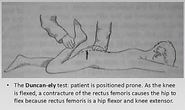
elys test

Positive test:

When the patient is unable to flex or touch the heels towards the buttocks and the hip of the affected side rises up from the table, the patient will also feel or complain of pain or tingling in the back or legs indicates a positive Ely’s test.

  • Facebook
  • Twitter
Clinical Skills and Approaches Clinical examinationMusculoskeletal systemOrthopedics

Post navigation

Previous post
Next post

Related Posts

Clinical Skills and Approaches lhermittes sign

Lhermitte’s Sign

Jul 11, 2016Jul 11, 2016

Synonym: Lhermitte’s phenomenon, Barber chair phenomenon Definition of Lhermitte’s Sign Lhermitte’s sign (pronounced Ler-meets) is an electric shock-like sensations, spreading along the spine in a cervico-caudal direction and into the arms and legs felt upon active or passive flexion of the neck. Reverse Lhermitte’s Sign is defined when symptoms are…

Read More
Clinical Skills and Approaches Bryant's triangle

Bryant’s triangle (Examination of Hip)

Jun 4, 2014Sep 5, 2022

Synonyms: Iliofemoral triangle Position: The patient lies supine with the pelvis square, and the limbs in identical position. Points of the Bryant’s triangle: The Bryant’s triangle is a right angled formed by: Anterior Superior Iliac Spine (ASIS) Tip of Greater trochanter Junction of perpendiculars from the 2 points above (1st draw…

Read More
Clinical Skills and Approaches renal angle tenderness

Costovertebral or Renal angle tenderness

Jul 26, 2019Jul 26, 2019

Synonyms: Murphy’s kidney punch, CVA tenderness (CVAT) Anatomy of Costovertebral angle or Renal angle: Costovertebral angle is formed by the junction of the 12th, or lowermost, rib with the paravertebral muscles, which run parallel to and on both sides of the vertebral column. Eliciting costovertebral angle tenderness: Position of patient:…

Read More

Leave a Reply Cancel reply

Your email address will not be published. Required fields are marked *

This site uses Akismet to reduce spam. Learn how your comment data is processed.

Pre-clinical (Basic Sciences)

Anatomy

Biochemistry

Community medicine (PSM)

Embryology

Microbiology

Pathology

Pharmacology

Physiology

Clinical Sciences

Anesthesia

Dermatology

Emergency medicine

Forensic

Internal medicine

Gynecology & Obstetrics

Oncology

Ophthalmology

Orthopedics

Otorhinolaryngology (ENT)

Pediatrics

Psychiatry

Radiology

Surgery

RSS Ask Epomedicine

  • What to study for Clinical examination in Orthopedics?
  • What is the mechanism of AVNRT?

Epomedicine weekly

  • About Epomedicine
  • Contact Us
  • Author Guidelines
  • Submit Article
  • Editorial Board
  • USMLE
  • MRCS
  • Thesis
©2026 Epomedicine | WordPress Theme by SuperbThemes