Skip to content
Epomedicine

Mnemonics, Simplified Concepts & Thoughts

Epomedicine

Mnemonics, Simplified Concepts & Thoughts

Case of Cyanotic Congenital Heart Disease : PGE1 saves life

Dr. Pedchrome, Jun 2, 2020Jun 2, 2020

A Single Male baby was born at 38 weeks of gestation with birth weight of 3.1 kg through Normal vaginal delivery. At birth the child had cried immediately. At 15 minutes of life, the child developed central cyanosis. There was no respiratory distress and heart rate was normal range.

Child was shifted immediately to NICU. Saturation was 70% and which reached upto 84% with oxygen supplementation. On auscultation, chest was unremarkable but on cardiac auscultation, a faint murmur could be heard. Hyperoxia test was also performed and the baby failed the hyperoxia test.

Fetal Circulation Made Easy

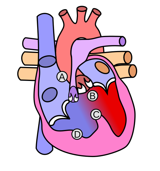
Immediately echocardiographic screening was done to look for possible presence of a duct dependent circulation. Echocardiography showed large VSD (Ventricular septal defect) with over-riding of Aorta with severe pulmonary stenosis. PDA was seen with left to right shunt measuring 3mm. It was case of TOF (Tetralogy of Fallot) with PDA (Patent Ductus Arteriosus) – a ductus dependent circulation as the baby had severe PS (Pulmonary Stenosis).

Tetralogy of Fallot
A – Pulmonary stenosis; B – Overriding of aorta; C – VSD; D – RVH
Wapcaplet / CC BY-SA

Chest Xray showed oligemic lung fields with upturned apex of the cardiac silhouette.

Patent ductus arteriosus
NIH / Public domain

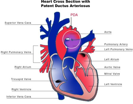
In any scenario, Prostin (PGE1) would have been started but since echocardiography showed patent PDA, it was withheld and monitoring was planned. On the second day, the baby was deteriorating, saturation was not maintained and reached as low as 60%. In general, SpO2 above 75% is adequate for Cyanotic heart diseases. Echocardiography was reassessed and the PDA was still patent at 2-3mm.  The desaturation was not explained by CHD. So, the child was kept under CPAP and finally intubated for the drop in saturation.

After intubation, the SpO2 worsened to 30-40%. Since the cardiologist was at the spot, reassessment was done with Echocardiography. PDA was patent with 2 mm size. A trial of PGE1 (Prostin) was given. After 5 minutes, SpO2 improved to 60%, heart rate improved from 100 to 120 and by 10 minutes SpO2 was 87% under MV and the baby looked much better.

Retrospective analysis:

It was a case of a closure or narrowing of PDA in a duct dependent circulation. The child was saved by PGE1 which keeps the PDA open, until the definitive management – BTS (Blalock Taussig Shunt).

A PDA connects the proximal left pulmonary artery to the descending aorta just distal to the left subclavian artery and is important in the foetal circulation as it diverts blood flow from the right ventricle to the descending aorta.

Pediatric doctor
Dr. Pedchrome

MD Pediatrics and Fellowship Neonatology, he chooses to stay anonymous. He often writes his views online as well as share few important topics for medical students, doctors and specially parents. He does research in pediatrics.

11 shares
  • Facebook11
  • Twitter
Case Reports Cardiovascular systemNeonatesPediatrics

Post navigation

Previous post
Next post

Related Posts

Case Reports

A case of EA/TOF (esophageal atresia/tracheoesophageal fistula)

Jan 26, 2024Jan 26, 2024

Case summary A female neonate was born via emergency cesarean section at 36 weeks of gestation. The indication of LSCS was polyhydramnios with fetal bradycardia. The birth weight of the neonate was 1700 grams following which the baby was shifted to NICU for observation. I, as a surgical resident was…

Read More
Case Reports

Mohr Syndrome (OFD2): An Orthopedic Insight from a Rare Case

Nov 9, 2025Nov 23, 2025

Introduction Mohr syndrome, or Orofaciodigital syndrome type II (OFD2), represents a very rare genetic disorder in which there is a triad of craniofacial anomalies, oral malformations, and digital deformities. While classic findings such as lobulated tongue, cleft palate, and broad nasal root mainly remain in the domain of pediatricians and…

Read More
Case Reports adh release

Craniopharyngioma – Case Discussion

Jan 20, 2016Jun 12, 2016

Case Summary A 38 year old female came with: Headache for 6 months Diminution of peripheral vision bilatetrally Irregular menstrual cycle for the last 1 year Now, polydypsia and excessive urination Her prolactin level was 115.6 ng/ml (increased), FSH was 1.3 mIU/ml (decreased) and LH was 0.242 mIU/ml (decreased). CT…

Read More

Leave a Reply Cancel reply

Your email address will not be published. Required fields are marked *

This site uses Akismet to reduce spam. Learn how your comment data is processed.

Pedchrome. Case of Cyanotic Congenital Heart Disease : PGE1 saves life [Internet]. Epomedicine; 2020 Jun 2 [cited 2025 Dec 20]. Available from: https://epomedicine.com/clinical-cases/cyanotic-congenital-heart-disease-pge1/.

Pre-clinical (Basic Sciences)

Anatomy

Biochemistry

Community medicine (PSM)

Embryology

Microbiology

Pathology

Pharmacology

Physiology

Clinical Sciences

Anesthesia

Dermatology

Emergency medicine

Forensic

Internal medicine

Gynecology & Obstetrics

Oncology

Ophthalmology

Orthopedics

Otorhinolaryngology (ENT)

Pediatrics

Psychiatry

Radiology

Surgery

RSS Ask Epomedicine

  • What to study for Clinical examination in Orthopedics?
  • What is the mechanism of AVNRT?

Epomedicine weekly

  • About Epomedicine
  • Contact Us
  • Author Guidelines
  • Submit Article
  • Editorial Board
  • USMLE
  • MRCS
  • Thesis
©2025 Epomedicine . All rights reserved.