Bidhan Sigdel, Sulabh Shrestha, Amrit Devkota
Fourth Year MBBS, KISTMCTH
ABSTRACT
Basal Cell Adenoma (BCA) accounts for 1-2% of salivary gland tumor 1.BCA is rare benign epitheloid neoplasm of salivary gland, which was first described and documented as distinct clinical and pathological entity by Kleinsasser and Klein in 1967 and was recognized by WHO in 1991 as a histologically distinct entity 2. In the parotid, BCA is occasionally difficult to distinguish from pleomorphic adenoma due to its overlapping clinical and cytological features. We report a case of BCA which was initially treated as a case of pleomorphic adenoma, review literature and discuss the differential diagnosis of similar case.
KEY WORDS
Basal cell adenoma; FNAC; Pleomorphic adenoma; Superficial parotidectomy
INTRODUCTION
Basal cell adenoma is a rare benign neoplasm of the salivary glands that derives its name from the basaloid appearance of the tumor cells. It preferentially occurs in the parotid gland during the sixth and seventh decades of life. The clinical presentation most frequently seen is a slow-growing, asymptomatic, movable, round or oval mass measuring less than 3.0 cm in diameter, encapsulated and well circumscribed. Histologically, BCA is composed of basaloid cells with eosinophilic cytoplasm, indistinct cell border, sharply delineated from the stroma by basement membrane, without mesenchymal component distributed in solid, trabecular, tubular or membranous pattern and may show cystic changes and squamous differentiation.
CASE REPORT
A 66 year old gentleman presented to Department of Surgery, KISTMCTH, with painless, slowly progressing swelling in left parotid region for 5 years. His other medical history was normal except his smoking habit for 30 years.
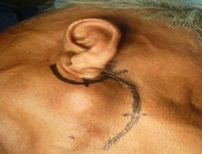
On examination, it was 4X4 cm2, well defined, smooth, firm to hard, non fluctuant, mobile swelling in left parotid region which had raised the ear lobule on the affected side strongly indicative of parotid enlargement. There were no signs of facial nerve palsy. Other ENT and systemic examination were normal.
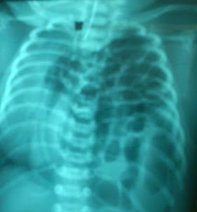
With the suspected diagnosis of superficial left parotid gland tumor, USG was performed which revealed 4*2cm2 complex cystic mass probably benign in nature without any lymph node involvement. Color Doppler showed minimal vascularity within the solid component. Then FNAC was done which showed congested scattered foamy macrophage and occasional fragments of uniform stromal cell with elongated nuclei, inconspicuous nucleoli and scanty poor cytoplasm, suggesting benign cystic neoplasm.
On the basis of these data, provisional diagnosis of pleomorphic adenoma was made and patient underwent superficial parotidectomy.
Through pre-auricular incision, superficial layer of parotid gland was removed and the mass was excised. Facial nerve was identified and was conserved during the operation.
Operative finding shows well capsulated heterogeneous mass with dirty blackish brown fluid coming out from superficial lobe of parotid.
Histology show tumor cells arranged in trabecular, tubular and some showing ductal pattern. The cells show mild pleomorphism with moderate amount of cytoplasm and nucleus with inconspicuous nucleoli and are frequently intervened by eosinophilic hyalinized stroma. Some of the areas shows tumor cell with squamous differentiation. Lymphovascular/perineural invasion was not noted. The histological study confirmed diagnosis as basal cell adenoma of parotid gland.
In post operative period, patient developed mild left facial nerve palsy which was gradually improving on following days.
DISCUSSION
Parotid gland, the largest salivary gland, is surrounded by continuation of investing layer of cervical fascia and has two lobes arbitrarily divided by facial nerve: Superficial lobe (70%) that lies over the posterior part of the ramus of mandible and Deep lobe (30%) that lies behind the mandible and medial pterygoid muscle.
Salivary gland tumor represents less than 3%of all tumors in general population. Approximately 88% of salivary gland neoplasms are of epithelial origin and benign adenoma accounts for about 66% of salivary tumor. Basal Cell Adenoma (BCA) accounts for 1-2% of salivary gland tumor 3.
BCA is rare benign epitheloid neoplasm of salivary gland, which was first described and documented as distinct clinical and pathological entity by Kleinsasser and Klein in 1967. In 1991, WHO recognized BCA as histologically distinct entity and in 2005 it was classified as 1 of 9 subcategories of salivary gland epithelial tumor 4.WHO defines BCA as “A distinctive benign neoplasm composed of basaloid cells organized with prominent basal cell layer and distinct basement membrane like structure and no myxochondral stroma as seen in pleomorphic adenoma.” 5
BCA arises from epithelial cell and earliest change seen is hyperplasia of basal cell of striated ducts. Continuous proliferation of the ductal cells leads to formation of microadenoma which eventually develops as adenoma.
BCA exclusively occurs in adults in 5th – 7th decade of life with the average age of 57 years with slight female predominance (M:F=1:2) 6. Clinically, tumor presents as painless slowly growing asymptomatic freely mobile firm mass most commonly in superficial lobe of parotid (80%),as in case reported above. However, BCA can occur in unusual sites such as upper and lower lip, buccal mucosa, palate and nasal septum. Facial pain and facial nerve palsy are extremely rare presentation of this tumor. This clinical presentation is similar with pleomorphic adenoma making BCA difficult to diagnose on basis of clinical examination alone.
Differential diagnosis must be established with other salivary gland neoplasm such as pleomorphic adenoma, adenoid cystic carcinoma, basal cell adenocarcinoma. The monomorphic appearance and absence of chondroid tissue and myxoid stroma differentiate BCA from pleomorphic adenoma. Tendency of perineural invasion even in early stage distinguish adenoid cystic carcinoma from BCA. Basal cell adenocarcinoma shows an infiltrative growth, more mitotic figure (>4 mitotic count/10HPF) as compared to BCA. Apart from these conditions it is important to consider other benign lesions like mucocele, sebaceous cyst, lipoma, nasolabial cyst, parotid abscess in differential diagnosis.
Diagnostic imaging for conclusion of suspected neoplastic disease in salivary gland includes sonography, CT scan, CT sialography and MRI. Sonography is often the initial investigation in parotid gland mass as done in our case because of its easy availability, low cost, non invasive technique and capable of guided biopsy. CT and MRI are better than sonography for assessing full tumor extent as they will confirm the mass being investigated originated from the gland or not and provides diagnostic information such as size, margin, invasive nature of tumor and its relation with surrounding structures. We acknowledge our limitation in not performing CT scan because of our patient’s socio economic status. We might have diagnosed the condition with CT scan preoperatively rather than misdiagnosing as pleomorphic adenoma.
FNAC can be used as both diagnostic and screening test. FNAC is useful in establishing whether a given lesion is inflammatory or neoplastic or lymphoma or an epithelial tumor or metastatic tumor. However, a specific diagnosis through FNAC can only be made in 60-75%of cases 7. Cytologically, the distinction between pleomorphic adenoma and BCA is difficult due to overlapping morphological features and sometimes because of inadequate sampling of tissue. FNAC can be reliable and specific diagnosis is often possible but it is advised to perform tissue biopsy for histological examination for confirmation of diagnosis, as done in our case.
Primary treatment of BCA is surgical excision by means of Superficial Parotidectomy or Total Parotidectomy in cases of membranous variant of BCA. Recurrence rate for solid and trabecular-tubular is almost non-existent but membranous type have high recurrence rate (24%)8.Although rare, malignant transformation is common in membranous type as compared with solid and trabecular-tubular type.
Complication of treatment includes temporary facial nerve paralysis( approximately in 30% of operation but recovers rapidly within 6 weeks) except when branches of facial nerve have been deliberately sacrificed. Frey’s syndrome is a regular sequale of parotidectomy occurring in up to 54% of cases 9. Anesthesia of the ear lobe can occur if great auricular nerve is damaged. Other rare complications such as siaocele or salivary fistula occasionally follow parotidectomy.
CONCLUSION
BCA, being rare tumor, is difficult to diagnose only by means of clinical judgment and imaging modalities. FNAC is usually reliable and specific but can sometimes be misleading. The final diagnosis requires complete histopathological examination. It is equally important to consider malignant counterpart and other benign lesion in differential diagnosis of BCA. Superficial parotidectomy with excision of tumor mass is treatment of choice in benign salivary neoplasm including BCA. Even though BCA is benign condition with good prognosis, it is advised for long term follow up in order to detect recurrence in prompt time.
ACKNOWLEDGEMENTS
Department of Surgery, KIST Medical College, Imadol, Lalitpur
Department of Pathology, KIST Medical College, Imadol, Lalitpur
REFERENCES
- González-García R, Nam-Cha SH, Muñoz-Guerra MF, Gamallo-Amat C. Basal cell adenoma of the parotid gland. Case report and review of the literature. Med Oral Patol Oral Cir Bucal 2006;11:E206-9.
- Irem Paker, M.D., Demet Yilmazer, M.D., Ata Turker Arikok, M.D.,Guleser Saylam, M.D., and Sema Hucumenoglu, M.D.Basal Cell Adenoma WithExtensive Squamous Metaplasia and Cellular Atypia:A Case Report With Cytohistopathological Correlation and Review of the Literature Diagnostic Cytopathology, Vol 40, No 1
- Mijung Jang, Dongwoo Park, Seung Ro Lee, Chang Kok Hahm, Youngsun Kim, Yongsoo Kim,Choong Ki Park, Kyung Tae, Moon Hyang Park, and Yong Wook Park Basal Cell Adenoma in the Parotid Gland: CT and MR Findings AJNR Am J Neuroradiol 25:631–635, April 2004
- Chan-Woo Kim, Seong-Gon Kim Basal cell adenoma misdiagnosed as an adenoid cystic carcinoma in the parotid gland (J Korean Assoc Oral Maxillofac Surg 2012;38:314-7)
- Russell R.C.G, Williams N.S, Bulstrode C.J.K, Bailey’s and Love’s Short Practice of Surgery, 23rd edition
- WHO Classification of Tumors, Volume 9 IARC WHO Classification of Tumors, No 9 Barnes, L., Eveson, J.W., Reichart, P., Sidransky, D. IARC
- L. Shi Y.-X.J. Wang C. Yu F. Zhao P.-D. Kuang G.-L. Shao CT and Ultrasound Features of Basal Cell Adenoma of the Parotid Gland: A Report of 22 Cases with Pathologic Correlation