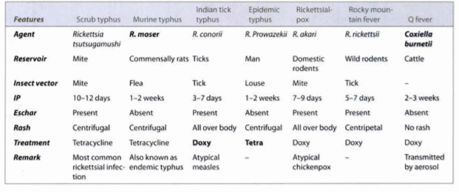
Important features of Rickettsia and Rickettsial Diseases Obligate intracellular parasites like Chlamydia – can survive only in host cells Cannot produce their own ATP, so they utilize the ATP of a host cell Gram negative coccobacillia, and short bacilli that grow strictly in eukaryotic cells (unable to grow in cell…
Author: Epomedicine
PGMEE, MRCS, USMLE, MBBS, MD/MS
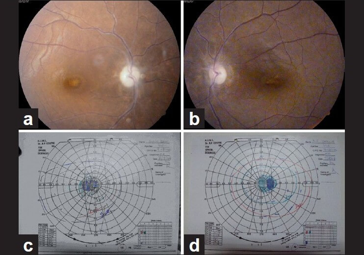
Ethambutol Induced Optic Neuropathy
Mechanism of Ethambutol induced optic neuropathy Ethambutol is metabolized to a chelating agent. The chelating agent formed then may impair the function of metal-containing mitochondiral enzymes, such as the copper containing cytochrome-c oxidase of complex IV and the iron containing NADH:Q oxidoreductase of complex I. These mitochondrial respiratory chain play…
Blog
Human Growth Hormone – Elixir of youth?
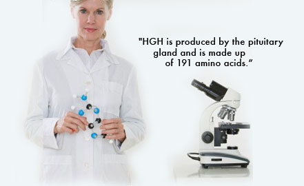
Human Growth Hormone (HGH) also known as somatotropin is synthesized and secreted by the acidophilic cells of pituitary gland in response to Growth hormone releasing hormone (GHRH). HGH is a protein (peptide) hormone composed of a chain of 191 amino acids. HGH production is inhibited by another hormone known as…
PGMEE, MRCS, USMLE, MBBS, MD/MS
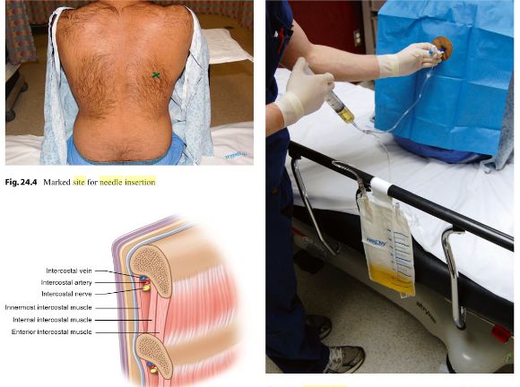
Thoracocentesis : Practical Essentials
Absolute contraindications of thoracocentesis There are no absolute contraindications to diagnostic thoracocentesis. If clinical judgement dictates that the information gained from the pleural fluid analysis may help in diagnosis and therapy, thoracocentesis should be performed. Necessity of an immediate alternative procedure such as open thoracostomy or thoracotomy is the only…
PGMEE, MRCS, USMLE, MBBS, MD/MS
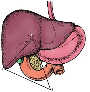
Gastrinoma (Passaro’s) triangle
Boundaries of Gastrinoma Triangle It is an imaginary triangle formed by 3 points in upper abdomen: Apex: Cystic duct-CBD junction Inferior point: 2nd part duodenum-3rd part duodenum junction Left lateral point: Pancreatic neck-body junction This area is located to the right of Superior Mesenteric Artery (SMA) i.e. Proximal duodenum and…
PGMEE, MRCS, USMLE, MBBS, MD/MS
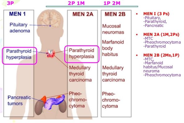
MEN syndrome : Mnemonics
MEN syndrome is an autosomal dominant (AD) predisposition to developing multiple endocrine tumors. Mnemonic: MEN are Dominant. Points to remember: MEN I or MEN 1 (Wermer’s syndrome) Mnemonic: 3 X P’s MEN IIA or MEN 2A (Sipple’s syndrome) Mnemonic: 3 X C’s MEN IIB or 2B (MEN III) Mnemonic: 2C 2M…
PGMEE, MRCS, USMLE, MBBS, MD/MS
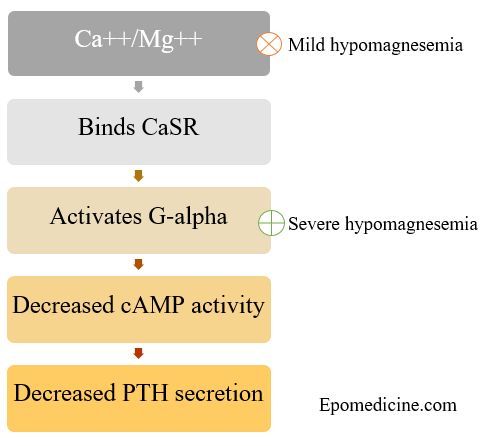
Why mild hypomagnesemia causes hyperparathyroidism and severe hypomagnesemia causes hypoparathyroidism?
You must have seen the statement in First Aid that says – low serum magnesium causes increase in Parathyroid hormone secretion and very low serum magnesium causes decrease in Parathyroid hormone secretion. Doesn’t this make you curious? Let’s explore the underlying mechanism in depth. How Calcium and Magnesium Mediated PTH…
PGMEE, MRCS, USMLE, MBBS, MD/MS
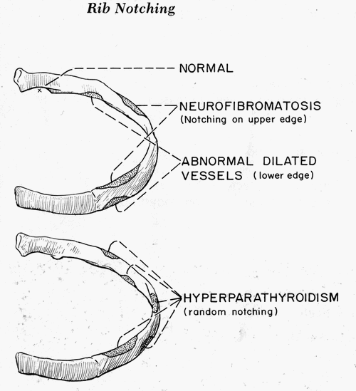
Rib Notching
Normal Rib Notching A small notch near the costo-vertebral joint is normal, so pathologic rib notching is more likely if the notching is more lateral. Types of Pathological Rib Notching 1. Superior rib notching 2. Inferior rib notching (more common) Inferior Rib Notching (Roesler’s sign) Mechanism: Enlargement of one or…