Synonym: Trichobateriosis axillaris, Bacterial trichonodosis, Trychomycosis palmellina
Definition: Asymptomatic superficial bacterial infection (mycosis is a misnomer because it is not a fungal infection) primarily of the axillary hairs (can also occur in pubic, perianal and scalp hair) caused by Corynebacterium sp.
Causative organism: Corynebacterium species particularly Corynebacterium flavescens
Mode of infection:
- Bacteria comes in contact with the hair shaft, and adhere to the surface, or the cuticle, of the hair, using a cement-like substance that is insoluble in water
- Micro-organism does not penetrate to the medulla’s cortex of the hair; instead, it only adheres strongly to the surface of the hair and develops slowly until it forms concretions around the hair shaft
- Encapsulated corynebacteria entrapped in a biofilm serve as the adhering mechanism, which possibly helps the organism to escape immunological attack by the host
Clinical variants:
- Trichomycosis flava (yellow)
- Trichomycosis rubra (red)
- Trichomycosis nigra (black)
Predisposing factors:
- Warm and humid climate
- Excessive sweating
- Inadequate hygiene
- Not shaving the area (hence, more common in male than female)
- Young adults
Clinical features:
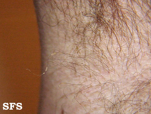
- Formation of concretions around the hair shaft:
- Early: Invisible and only a slight thickening can be “felt” on palpation and later the bacterial masses remain isolated or independent (pediculosis-like)
- Chronic: Concretions extend along the entire length of the hair until they form a sheath, causing the hair to thicken, turn a yellowish, red or black color, and become creamy, opaque and soft.
- Bromhidrosis (malodor)
- Hyperhidrosis (excessive sweating)
- May stain clothing
Wood’s lamp examination: Pale-yellow fluorescence
Dermoscopy: Yellowish-white masses with a waxy appearance, adherent to the hair (feather, brush or skewer sign); Concretions with the appearance of a rosary of crystalline stones; a flame-like pale yellowish adherent nodule (plume sign)
KOH examination: Mucoid sheath around hairs
Gram stain: Hair shafts colonized by gram-positive coccobacilli
Culture: A mixture of microorganisms may be identified: Corynebacterium sp. (white-yellow growth) and Serratia mascescens (red growth)
Treatment:
A. General measures:
- Adequate hygiene
- Shaving of the affected area for 2-3 weeks
- Control of hyperhidrosis
B. Cleansing methods: Use of sulfur soaps
C. Topical treatments:
- 3% sulfur
- 2% sodium hypochlorite
- 1% mercury chloride
- 5% Benzoyl peroxide
- Antibiotics: Fusidic acid, 2% Erythromycin, 1% Clindamycin
- Antimycotic agents: Naftifine, Some azole derivatives
References:
- Bonifaz A, Váquez-González D, Fierro L, Araiza J, Ponce RM. Trichomycosis (trichobacteriosis): clinical and microbiological experience with 56 cases. Int J Trichology. 2013 Jan;5(1):12-6. doi: 10.4103/0974-7753.114704. PMID: 23960390; PMCID: PMC3746219.
- https://www.actasdermo.org/en-trichomycosis-axillaris-clinical-wood-lamp-articulo-S1578219017300331
- https://escholarship.org/uc/item/7488g7vb