Skip to content
Epomedicine

Mnemonics, Simplified Concepts & Thoughts

Epomedicine

Mnemonics, Simplified Concepts & Thoughts

Superficial Cervical plexus

Superficial Cervical Plexus Block : Mnemonic

Epomedicine, Nov 3, 2015

This is a visual mnemonic for the nerve arrangement of superficial cervical cutaneous branches of cervical plexus. This mnemonic was created only for the ease to remember and may not resemble exact anatomy.

Superficial Cervical plexus

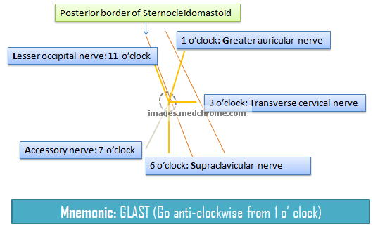
The site of injection for superior cervical plexus nerve block is the midpoint of posterior border of sternocleidomastoid. Assuming this as a center point, the various position of nerves, from north going anti-clockwise is given by mnemonic “GLAST”:

  1. 1 o’ clock: Greater auricular nerve (C2,C3) – Innervates skin over the parotid gland, angle of jaw and posterior ear
  2. 11 o’ clock: Lesser occipital nerve (C2) – Innervates scalp behind and above ear
  3. 7 o’ clock: Spinal accessory nerve (XI) – This doesn’t belong to superficial cervical plexus and lies deep to the sternocleidomastoid muscle and innervates sternocleidomastoid and trapezius
  4. 6 o’ clock: Supraclavicular nerve (C3,C4) – Divides into medial, intermediate and lateral branches and supply sensation over shoulder, lateral neck and anterior upper thoracic wall
  5. 3 o’ clock: Transverse cervical nerve (C2, C3) – Innervates skin of front and side of neck (anterior triangle)

All these nerves are also the contents of the occipital triangle of neck. Except spinal accessory nerve, all these nerves crosses superficial to the sternoclediomastoid muscle. In general, the superficial cervical plexus supply the skin of anterolateral neck. Superficial cervical plexus block is indicated for skin surgeries of head and neck.

Origin of cervical plexus: anterior rami of the C1 – C4 nerve roots

Location of cervical plexus: anterior to the cervical vertebrae and posterior to the sternocleidomastoid muscle.

Components of cervical plexus:

  1. Cutaneous branches, which supply the lesser occipital, greater auricular, transverse cervical, and supraclavicular nerves
  2. Ansa cervicalis, which innervates the infrahyoid and geniohyoid muscles
  3. Phrenic nerve, which is the only motor nerve to innervate the diaphragm
  4. Contributions to the accessory nerve (CN XI), which innervates the sternocleidomastoid and trapezius muscles
  5. Direct muscular branches, which supply prevertebral muscles of the neck.

Procedure:

Insert the needle ( 22-gauge, 5-cm, short bevel needle) at the midpoint of the posterior border of the sternocleido – mastoid muscle to approximately half the depth of the muscle, and inject 3 to 4 mL of local anesthetic. Also perform a subcutaneous injection of additional local anesthetic cephalad and caudad along the length of the sternocleidomastoid muscle posterior border.

8 shares
  • Facebook8
  • Twitter
PGMEE, MRCS, USMLE, MBBS, MD/MS AnatomyAnesthesia

Post navigation

Previous post
Next post

Related Posts

PGMEE, MRCS, USMLE, MBBS, MD/MS intercellular junctions

Cell-Cell or Interceullular Junctions

May 22, 2019May 22, 2019

Junction Location Composition Function Remarks Tight junction (Zonula occludens) Epithelial cell-Epithelial cell Claudin and Occludin (attaches to cytoskeleton – actin filament) Prevents paracellular movement of solutes and diffusion Adherens junction (Zonula adherens) Epithelial cell-Epithelial cell E-cadherin (attaches to cytoskeleton – actin filament) Belt desmosome (Anchoring) Cadherins are Ca2+ depndend adhesion…

Read More
PGMEE, MRCS, USMLE, MBBS, MD/MS

Inferior Venacava (IVC) Tributaries and Abdominal Aorta Branches : Mnemonic

May 11, 2024May 11, 2024

Inferior Venacava (IVC) Tributaries Mnemonic: I Like To Rise So Incredibly High 1. common Iliac veins (L5) – Origin or Formation 2. Lumbar veins (L1-L5) 3. Testicular/gonadal veins – right (L2) 4. Renal veins (L1) 5. Suprarenal veins – right (L1) 6. Inferior phrenic veins (T8) 7. Hepatic veins (T8)…

Read More
PGMEE, MRCS, USMLE, MBBS, MD/MS palatogenesis

Face and Palate Development – Embryology made Easy

Aug 7, 2016Aug 7, 2016

4 week embryo 5 mesenchymal prominences (facial primordia) appear in relation to the stomodeum (a depression in the surface ectoderm which marks the future mouth and oral cavity): Cranially: Frontonasal prominence (unpaired) Laterally: Maxillary prominence (paired; 1st pharyngeal arch) Caudally: Mandibular prominence (paired; 1st pharyngeal arch) 5 week embryo Localized…

Read More

Leave a Reply Cancel reply

Your email address will not be published. Required fields are marked *

This site uses Akismet to reduce spam. Learn how your comment data is processed.

Epomedicine. Superficial Cervical Plexus Block : Mnemonic [Internet]. Epomedicine; 2015 Nov 3 [cited 2025 Dec 23]. Available from: https://epomedicine.com/medical-students/superficial-cervical-plexus-block-mnemonic/.

Pre-clinical (Basic Sciences)

Anatomy

Biochemistry

Community medicine (PSM)

Embryology

Microbiology

Pathology

Pharmacology

Physiology

Clinical Sciences

Anesthesia

Dermatology

Emergency medicine

Forensic

Internal medicine

Gynecology & Obstetrics

Oncology

Ophthalmology

Orthopedics

Otorhinolaryngology (ENT)

Pediatrics

Psychiatry

Radiology

Surgery

RSS Ask Epomedicine

  • What to study for Clinical examination in Orthopedics?
  • What is the mechanism of AVNRT?

Epomedicine weekly

  • About Epomedicine
  • Contact Us
  • Author Guidelines
  • Submit Article
  • Editorial Board
  • USMLE
  • MRCS
  • Thesis
©2025 Epomedicine . All rights reserved.