Skip to content
Epomedicine

Mnemonics, Simplified Concepts & Thoughts

Epomedicine

Mnemonics, Simplified Concepts & Thoughts

cholesterolosis of gallbladder

Strawberry Gallbladder (Cholesterolosis)

Epomedicine, Jun 12, 2015Aug 17, 2015
strawberry gallbladder
Strawberry gallbladder noted incidentally in a cholecystectomy specimen for cholelithiasis.

Synonyms:

Strawberry gallbladder is also known as hyperplastic cholecystoses. Hyperplastic cholecystoses are a spectrum of non-neoplastic proliferative disorders caused by deposition of cholesterol-laden macrophages within the wall of the gall bladder. The cholecystose range from abnormalities of the gallbladder wall (adenomyomatosis and strawberry gall bladder) to gallbladder polyps extending into the lumen.

Definition: 

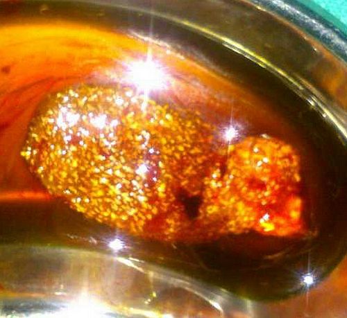
Cholesterolosis occurs due to the accumulation of cholesterol esters and triglycerides in subepithelial macrophages and gallbladder epithelium. It is associated with bile supersaturation with cholesterol, but not with increased serum cholesterol. It is usually asymptomatic and detected incidentally in 30-50 % of cholecystectomy specimens. Cholesterolosis occurs in 2 major forms: localized and diffuse. Strawberry gallbladder refers to diffuse cholesterolosis characterized by tiny mural cholesterol deposits likened to strawberry seeds. It is a pathologic diagnosis rather than a clinical diagnosis.

Clinical features:

This is usually asymptomatic but sometimes may exhibit mild symptoms such as indigestion, gas and distress after meals. Pain in the upper quadrant is also noted.

Epidemiology:

In a Survey done to determine the association between cholesterolosis and gall bladder malignancy following results were observed :

  • Sample: 3123 cases
  • Gender predominance: more common in women than in men
  • Cholesterolosis has a strong negative association with gallbladder cancer

Gross features:

  1. Yellow, flat deposits on mucosal surface, focal or diffuse.
  2. May have speckled appearance (“strawberry gallbladder”), 20% are associated with cholesterol polyps.
  3. Polyps vary in size from 1-10 mm.
  4. Bile in the lumen is generally dark and thick, with a high concentration of cholesterol and sometimes floating yellow lipid particles (lipoidic corpuscles).

Microscopic features:

  1. Foamy macrophages in lamina propria and epithelium.
  2. Villous mucosal hyperplasia with macrophages at tips of villi.
  3. Usually no or minimal cholecystitis.
  4. Usually changes are restricted to gallbladder and don’t involve extrahepatic bile ducts.

Ultrasound features:

  1. Brightly echogenic non-mobile masses, with a ring down/ comet tail or reverberation artefact.
  2. No posterior acoustic shadowing.
  3. When cholesterosis is focal, it forms cholesterol polyps.

Primary concern:

Rule out gall bladder malignancy.

Submitted by: Dr. Sulabh Shrestha

3 shares
  • Facebook3
  • Twitter
PGMEE, MRCS, USMLE, MBBS, MD/MS Gastrointestinal systemGeneral SurgeryPathology

Post navigation

Previous post
Next post

Related Posts

PGMEE, MRCS, USMLE, MBBS, MD/MS concentration effect

Concentration effect, Second gas effect and Diffusion hypoxia

Oct 20, 2016Oct 20, 2016

Concentration effect The higher the concentration of an inhaled anesthetic, the faster the alveolar concentration approaches the inhaled concentration. This is referred to as concentration effect and is clinically significant only in cases where gases are administered in high concentration: Nitrous oxide Xenon Ostwald’s blood gas solubility coefficient: ratio of…

Read More
PGMEE, MRCS, USMLE, MBBS, MD/MS

Working formulation of NHL (Mnemonic)

Jan 16, 2019Feb 26, 2019

All follicular except large cell: Low grade All diffuse: Intermediate grade Blastic and non-cleaved: High grade

Read More
PGMEE, MRCS, USMLE, MBBS, MD/MS

Forearm muscles : Tricks to remember

Oct 28, 2015Oct 27, 2022

Anterior Forearm (Compartment) Muscles Total muscles: 8 (4 superficial + 1 intermediate + 3 deep) Mnemonic: Do it yourself as shown in the figure below! Place your thenar/hypothenar eminence over medial epicondyle and fan out 5 fingers with thumb resting below the 4 fingers. The 4 fingers represent superficial flexors…

Read More

Leave a Reply Cancel reply

Your email address will not be published. Required fields are marked *

This site uses Akismet to reduce spam. Learn how your comment data is processed.

Pre-clinical (Basic Sciences)

Anatomy

Biochemistry

Community medicine (PSM)

Embryology

Microbiology

Pathology

Pharmacology

Physiology

Clinical Sciences

Anesthesia

Dermatology

Emergency medicine

Forensic

Internal medicine

Gynecology & Obstetrics

Oncology

Ophthalmology

Orthopedics

Otorhinolaryngology (ENT)

Pediatrics

Psychiatry

Radiology

Surgery

RSS Ask Epomedicine

  • What to study for Clinical examination in Orthopedics?
  • What is the mechanism of AVNRT?

Epomedicine weekly

  • About Epomedicine
  • Contact Us
  • Author Guidelines
  • Submit Article
  • Editorial Board
  • USMLE
  • MRCS
  • Thesis
©2026 Epomedicine | WordPress Theme by SuperbThemes