Definition: Abnormal scapular motion which may refer to –
- Abnormal static positioning – including prominence of its medial border or inferior angle
- Abnormal scapular movement – including early scapular elevation on arm elevation or rapid downward scapular motion during arm descend
Causes:
1. Primary: Due to abnormalities arising in Scapulo-thoracic articulation, periscapular muscles and nerves
- Neurological dysfunction: Long thoracic nerve, Spinal accessory nerve, Dorsal scapular nerve
- Osseous: Osteochondromas (scapula/ribs), Fractures (scapula/ribs), Thoracic spine scoliosis/kyphosis
- Soft tissue: Congenital muscle absence (Serratus anterior, Trapezius, Rhomboids), Traumatic muscle avulsion (Serratus anterior), Scapulo-thoracic bursitis
- Voluntary
2. Secondary: Response to shoulder abnormalities
- Shoulder pain: causes reflex spasm of peri-scapular muscles
- Glenohumeral stiffness: causes abnormal scapular motion (increased scapulo-thoracic motion, leading to scapular fatigue & winging) trying to maintain overall motion
- Deltoid fibrosis
Types:
It is categorized into three main types based on the prominence of specific areas of the scapula during movement.
- Type I (inferomedial scapular border prominence)
- Type II (entire medial border prominence)
- Type III (superomedial scapular border prominence)
Clinical symptoms:
- Shoulder weakness (esepcially on arm abduction)
- Reduced functional performance
- Abnormal prominence of scapula
- Reduced shoulder motion
- Pain (dull/heaviness/burning) in scapula, neck & arm area
- Muscle pain (due to spasm of overcompensating scapular muscles)
- Shoulder drooping (trapezius dysfunction) & shoulder asymmetry
- Feeling of abnormal movement in shoulder
- Peri-scapular muscle wasting
Clinical signs:
- Possible surgical scars (iatrogenic cause)
- Muscle wasting
- Muscular, bursa or bony tenderness
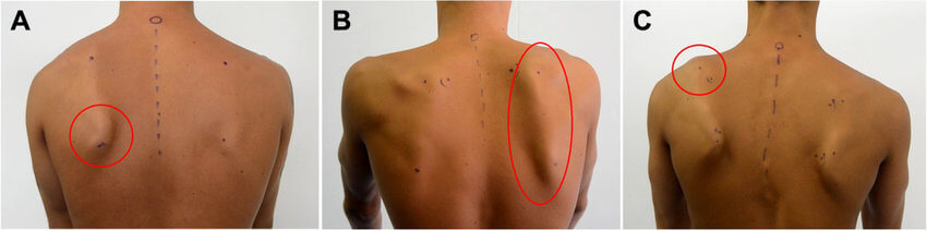
- Abnormal scapular posture – static and/or dynamic
- Winging:
- Static (pseudo-winging): due to structural abnormalities of scapula or ribs
- present at rest with arm by the side
- doesn’t increase on wall push-up
- Dynamic (true winging): due to muscle imbalance; increases on wall push-up
- Medial winging: Serratus anterior palsy
- Lateral winging: Trapezius or Rhomboid palsy
- Static (pseudo-winging): due to structural abnormalities of scapula or ribs
- Palpable crepitus
- Painful or limited shoulder movements
Investigations:
- Plain X-ray, CT: to look for osseous causes
- MRI: to look for osseous bursitis and soft tissue lesions
- Nerve conduction studies
- Electromyography
Management:
1. Secondary scapular dyskinesis: Manage the underlying primary shoulder disorder
2. Primary scapular dyskinesis:
a. Non-surgical:
- Leave alone
- Analgesia
- Steroid injection of bursae in scapulo-thoracic bursitis
- Physiotherapy:
- Maintain glenohumeral motion
- Stretch stiff muscles
- Improve posture
- Strengthen weak or compensatory muscles
- Correct muscle dis-coordination – biofeedback
b. Surgical:
- Nerve surgery: Neurolysis, Nerve grafting
- Muscle transfers:
- Trapezius dysfunction: Eden Lange procedure (transfer of Levator scapulae to acromion & Rhomboids to infraspinous fossa)
- Serratus anterior dysfunction: Transfer of sternocostal head of Pectoralis major with a graft extension (fascia lata, semitendinosus)
- Osseous procedures:
- Osteochondroma: Resection
- Malunions of scapular fractures: Osteotomy
- Scapulothoracic fusion (improves pain at the expense of motion)
SICK Scapula
The term SICK scapula was introduced to describe the pathological state of the scapula characterised by:
- Scapula malposition
- Inferior medial border prominence
- Coracoid pain and malposition
- Kinesis abnormalities of the scapula
This syndrome is often seen in overhead athletes and thought to contribute to the development of shoulder injuries. The scapula malposition consists of forward tilting and protraction (making it look “drooped”).
Reference: The Shoulder Made Easy – Charalambos Panayiotou Charalambous