Skip to content
Epomedicine

Mnemonics, Simplified Concepts & Thoughts

Epomedicine

Mnemonics, Simplified Concepts & Thoughts

Pterygium excsion bare sclera

Pterygium Excision

Epomedicine, Jun 2, 2014May 31, 2020

Indications for surgery:

1. Reduced vision secondary to:

  • Threatens visual axis: Pterygium advancing toward or already impinging visual axis
  • Induced astigmatism: Due to flattening of meridian of pterygium by fibrosis in atrophic stage

2. Cosmesis

3. Significant discomfort not relieved by medical therapy

4. Diplopia: Resulting from limited ocular motility secondary to muscle restriction

Surgical modalities:

1. Simple excision

2. Simple excision with bare sclera technique (90% recurrence)

3. Excision with adjunctive measures:

  • Postoperative Beta-radiation
  • Thiopeta drops
  • Intraoperative and Postoperative mitomycin C
  • Conjunctival autografting (5% recurrence)
  • Amniotic membrane autografting
  • Keratectomy and Lamellar keratoplasty: For recurrent recalcitrant pterygium

Procedure:

1. Anesthesia: Topical anesthesia

2. Preparation and draping: Cleanse eyelashes and lid margins with Povidone-iodine 5% on a cotton-tipped applicator and place 1-2 drops of povidone-iodine in fornix

3. Insert universal eye speculum

4. Forced duction test: To rule out any restriction of rectus muscle

5. Position eye with stay sutures and clamp

6. Demarcate the body of pterygium with cautery

Pterygium excsion bare sclera
A: Removal of head of pterygium
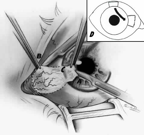
B: Removal of tail of pterygium
C: Removal of neck of pterygium
D: Bare sclera covered by conjunctival autograft

7. Remove head of pterygium (corneal portion): Lift and dissect off the cornea

8. Remove tail of pterygium (episcleral portion): Use Westcott scissors to cut along previously placed cautery marks; undermine pterygium with scissors and remove all pterygium and underlying tissue (including tenon’s capsule) down to bare sclera

9. Remove neck of pterygium (limbal portion): Using sharp and blunt dissection with scissors

10. Achieve hemostasis with the help of cautery

11. Polish the limbus with diamond burr to smoothen it

12. Next step differs depending upon the technique:

  • Simple excision: Conjunctiva is sutured back to cover the sclera
  • Bare sclera technique: Some part of conjunctiva is excised and its edges are sutured to the underlying episcleral tissue leaving some bare part of sclera
  • Conjunctival membrane autograft: Free conjunctiva from the same or opposite eye may be used as a graft to cover the bare sclera
  • Adjunct therapy with mitomycin C: Place Mitomycin C (MMC) soaked sponge onto the bare sclera for 2 minutes
  • Amniotic membrane graft: Amniotic membrane graft 1mm oversized on all sides is used to cover the bare sclera

13. Apply topical combination antibiotic and steroid ointment

14. Place light pressure patch and fox shield

  • Facebook
  • Twitter
Surgical Skills General SurgeryOphthalmology

Post navigation

Previous post
Next post

Related Posts

Surgical Skills

Intramedullary Nail Dynamization

Apr 29, 2025Apr 29, 2025

Dynamization of intramedullary nail is the process of converting a static interlocking nail (provides better fixation & rotational control) to a dynamic locking nail (allows axial loading of fracture to stimulate bone healing). Dynamization converts a bridging mode of fixation to the splinting mode. Timing of Dynamization: Dynamization should be…

Read More
Surgical Skills

Zancolli-Lasso Procedure

May 6, 2021May 6, 2021

Based on the preoperative clinical examination, the decision on which donor superficialis (FDS) tendon to use is made (commonly FDS middle finger is used). Transverse incision is made at the level of the distal palmer crease. Care should be taken to avoid the neurovascular bundles and the flexor tendons must…

Read More
Fellowship Blog

When Locking Screws Refuse to Budge – Techniques for Removing Jammed or Stripped Screws

Dec 6, 2025Dec 6, 2025

Written on August 10, 2025 And once again, today’s case reminded me that removing orthopedic implants can be more challenging than inserting them – especially when faced with a stubborn, stripped or jammed locking screw. We operated on a 25-year-old female with a united distal radius fracture, aiming to remove…

Read More

Leave a Reply Cancel reply

Your email address will not be published. Required fields are marked *

This site uses Akismet to reduce spam. Learn how your comment data is processed.

Epomedicine. Pterygium Excision [Internet]. Epomedicine; 2014 Jun 2 [cited 2026 May 14]. Available from: https://epomedicine.com/surgical-skills/pterygium-excision/.

Pre-clinical (Basic Sciences)

Anatomy

Biochemistry

Community medicine (PSM)

Embryology

Microbiology

Pathology

Pharmacology

Physiology

Clinical Sciences

Anesthesia

Dermatology

Emergency medicine

Forensic

Internal medicine

Gynecology & Obstetrics

Oncology

Ophthalmology

Orthopedics

Otorhinolaryngology (ENT)

Pediatrics

Psychiatry

Radiology

Surgery

RSS Ask Epomedicine

  • What to study for Clinical examination in Orthopedics?
  • What is the mechanism of AVNRT?

Epomedicine weekly

  • About Epomedicine
  • Contact Us
  • Author Guidelines
  • Submit Article
  • Editorial Board
  • USMLE
  • MRCS
  • Thesis
©2026 Epomedicine | WordPress Theme by SuperbThemes