Lytic and Sclerotic Bone Lesions
Mnemonic for Solitary lytic lesions: FEGNOMASHIC
- Fibrous dysplasia
- Enchondroma
- Eosinophilic granuloma
- Giant cell tumor
- Nonossifying fibroma
- Osteoblastoma, Osteosarcoma (telangiectatic)
- Metastases
- Myeloma
- Aneursymal bone cyst
- Simple bone cyst
- Hyperparathyroidism (brown tumor)
- Infection
- Chondroblastoma
- Chondromyxoid fibroma
Mnemonic for Multiple lytic lesions: FEMHI
- Fibrous dysplasia
- Eosinophilic granuloma/Enchondroma
- Metastasis/Mutiple myeloma
- Hyperparathyroidism (Browns tumor)/Hemangioma
- Infection
Mnemonic for Focal sclerotic bone lesions: HOME LIFE
- Healed non-ossifying fibroma
- Osteoma
- Metastases
- Ewing’s sarcoma
- Lymphoma
- Infection/Infarct
- Fibrous dysplasia
- Enchondroma
Mnemonic for Sclerotic Bone Metastases: 5 Bees Like Pollen
- Brain (Medulloblastoma, Meningiosarcoma)
- Bronchus (Primary lung carcinoma)
- Bowel (Carcinoid, Mucinous carcinomas)
- Bladder
- Breast (lytic, mixed or sclerotic)
- Lymphoma (more often lytic; Hodgkin’s lymphoma produces sclerotic lesions)
- Prostate (nearly always sclerotic)
Multiple lesions: 3 M
- Multiple myeloma
- Metastases
- Multiple bone (polyostotic) involvement: Brown tumors, Non-ossifying fibroma, Fibrous dysplasia, Multifocal osteomyelitis, Multifocal osteomyelitis, Langerhans cell histiocytosis
Location in bone
a. Epiphysis:
- Pediatric: Chondroblastoma
- Adult: Giant cell tumor (GCT) or Osteoclastoma, Clear cell chondrosarcoma
- Other differentials: Osteomyelitis, Paget disease
b. Metaphysis:
- Medullary:
- Simple (unicameral) bone cyst
- Aneurysmal (multicameral) bone cyst
- Enchondroma
- Fibrous dysplasia
- Chondromyxoid fibroma
- Conventional osteosarcoma
- Chondrosarcoma
- Pediatric GCT or osteocalstoma
- Osteochrondroma (most common benign bone tumor)
- Intra-cortical:
- Fibrous cortical defect (FCD)
- Osteo-fibrous dysplasia
- Osteoid osteoma
- Juxtacortical (Surface):
- Juxtacortical chondroma
- Periosteal osteosarcoma
- Paraosteal osteosarcoma
- Juxtacortical chondrosarcoma
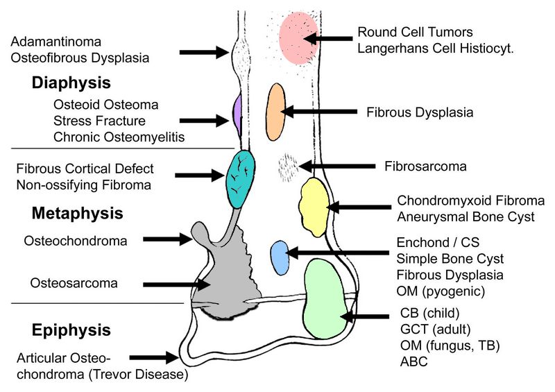
c. Diaphysis:
Mnemonic: FEMALE for Diaphyseal lucent lesions
- Fibrous Dysplasia (Fibrous bone tumor originating in diaphysis; “D” for dysplasia and dipahysis; all other fibrous tumors in metaphysis)
- Eosinophilic granuloma
- Metastasis/Myeloma
- Adamantinoma
- Lymphoma
- Ewing’s sarcoma and other round cell lesions
OR in general, remember diaphysis is AFORE metaphysis and epiphysis: Adamantinoma, Fibrous Dysplasia, Osteoma/Osteoblastoma, Round cell tumors, Endchondroma/Eosinophilic granuloma
- Medullary:
- Fibrous dysplasia
- Ewing sarcoma
- Endchondroma
- Cortical:
- Ossifying fibroma (Osseofibrous dysplasia)
- Osteoid osteoma (<2 cm; pain relieved by NSAIDs) and Osteoblastoma (>2 cm; pain not relieved by NSAIDs)
- Adamantinoma
Other medullary lesions which can occur in metaphysis or diaphysis
1. Localized Langerhans cell histiocytosis (LCH)
2. Myeloma
3. Metastases
4. Malignant vascular tumors
5. Lymphoma
2 Benign tumors that metastasize:
1. Giant cell tumor
2. Chondroblastoma
Location in Human body
a. Spine: Multiple myeloma, Metastatic tumors, Chordoma, Giant cell tumor, Chondrosarcoma, Osteoblastoma, Hemangioma
b. Phalanges: Enchondroma, Abscess, Metastatic tumors (especially lungs), Pigmented villonodular synovitis (PVNS), Giant cell tumor
c. Intra-articular: Synovial cell carcinomatosis, PVNS
- Adamantinoma: Tibia or Mandible (ameloblastoma)
- Osteoid osteoma: Proximal femur > Tibia > Posterior spine
- Osteoblastoma: Posterior spine > Long bones
- Osteoma: Facial bones
- Fibrous dysplasia: Proximal femur > Tibia , Ribs (most common site in mono-ostotic FD), Craniofacial skeleton
- Ewing’s sarcoma: Femur > Tibia
- Simple bone cyst: Proximal humerus > Proximal femur
- Aneurysmal bone cyst (ABC): Proximal femur > Tibia, Posterior spine, Pelvis
- Osteosarcoma: Distal femur > Proximal tibia
- Chondrosarcoma: Pelvis > Femur > Ribs > Humerus > Tibia
- Enchondroma: Small bones of hand and feet
- Osteochondroma (Exostosis): Distal femur
- Ivory osteoma (Eburnated osteoma): Frontal sinus
- Non-ossifying fibroma: Tibia
- Chondroblastoma: Distal femur and proximal tibia > Proximal humerus > Flat bones
- Giant cell tumor: Distal femur and proxima tibia
- Hemangioma: Anterior spine
- Myeloma: Anterior spine
- Glomus tumor: Tuft of phalanx
- Chordoma: Sacrum > Clivus > Anterior spine
- Intraosseous lipoma: Calcaneus
In vertebra:
1. Posterior part: Benign condition
2. Anterior part: Malignant condition (Exception: Hemangioma and Eosinophilic granuloma)
Patella, Calcaneus and most apophyses can be considered as “Epiphyseal equivalents” and hence, classic epiphyseal lesions like CB, GCT, ABC fall in the differentials.
Lucent lesions of sternum must be considered malignant unless proven otherwise.
Age group
a. <1: Neuroblastoma
b. 0-10: Ewing’s (tubular bones)
- Others: Solitary bone cyst (SBC)
c. 10-20: Osteosarcoma, Ewing’s (flat bones), Chondroblastoma, Osteoblastoma, Osteoid osteoma, Osteochondroma
- Others: SBC, ABC, NOF, Fibrous dysplasia, Chondromyxoid fibroma
d. 20-40: Giant cell tumor, Enchondroma, Osteoma, Osteoblastoma
e. >40: Metastatic, Myeloma, Chondrosarcoma, Paget’s disease
Mnemonic: SCAN Everything for Benign lytic lesions that rarely occur after 30 years of age –
1. Simple bone cyst
2. Chondroblastoma
3. Aneurysmal bone cyst
4. Non-ossifying fibroma
5. Eosinophilic granuloma
Mnemonic: MARCO for most common bone tumors over 30 years of age –
1. Metastatic tumors
2. Adult Round cell tumors (myeloma, lymphoma)
3. Chondrosarcoma
4. Osteosarcoma
Matrix
a. Chondroid matrix (Cartilage producing tumors): Small punctate or swirled areas of calcification (popcorn-like, curvilinear, speckled)
b. Osseous matrix (Bone producing tumors): Dense and confluent calcification (cloud-like, mashed potatoes)
c. Others (Fibrous dysplasia, Fibrosarcoma, Malignant fibrous histiocytoma, Solitary bone cyst): No or little calcification
Margins
Geographic lesion: If the tumor is not aggressive, bone gets time to lay itself down producing a distinct margin around the lesion. These produce bubbly lesions.
- Produced by benign lytic lesions (most of the FEGNOMASHIC described earlier)
Moth-eaten or Permeative lesion: If the tumor/disorder is aggressive, bone has not time to lay itself down producing ill-defined foci of lucency.
- Produced by aggressive infection or malignant tumor (Mnemonic: FIRE MD)
- Fibrosarcoma
- Infection (Osteomyelitis)
- Round cell tumors (Ewing’s, neuroblastoma, retinoblastoma)
- Eosinophilic granuloma
- Myeloma/Metastases
- Desmoid tumor
Periosteal reaction
- Solid or unilamellated: Non-aggressive lesion, periosteum has time to lay down thick bone (Osteoid osteoma)
- Multi-lamerllated or onion-skin: Intermediate aggressive lesion (Ewings)
- Spiculated, or “hair-on-end” (perpendicular to the cortex) or sunburst pattern: Most aggressive lesion (Malignancy)
Codman’s triangle: Elevation of periosteum away from the cortex. It may be seen in –
1. Conventional osteosarcoma
2. Ewing’s sarcoma
3. Osteomyelitis
4. Active aneurysmal bone cyst
5. Subperiosteal hematoma/abcess
In the end, we have a mnemonic for the headings under which, we describe bone tumors:
Mnemonic: CAMPS
- Cortical response
- Age (maturity of skeleton)
- Margins (transition zone) and Matrix
- Pattern of destruction and Periosteal reaction
- Site, Size and Soft tissue involvement