Skip to content
Epomedicine

Mnemonics, Simplified Concepts & Thoughts

Epomedicine

Mnemonics, Simplified Concepts & Thoughts

parinaud syndrome

Parinaud Syndrome – Anatomical Basis

Epomedicine, May 19, 2019May 19, 2019

Synonyms: Dorsal midbrain sydnrome, Pretectal syndrome

Main components of Parinaud syndrome:

  1. Vertical gaze palsy
  2. Convergence-retraction nystagmus
  3. Pupillary light-near dissociation
  4. Lid retraction (Collier’s sign)
Madhero88 [CC BY-SA 3.0], via Wikimedia Commons

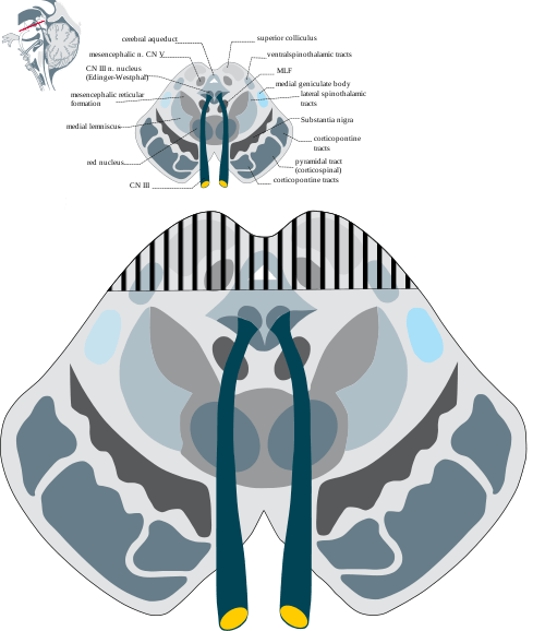
Anatomical basis of Parinaud syndrome:

Site of lesion: Dorsal midbrain

  • Close to cerebral aqueduct and pineal gland – hydrocephalus and pinealoma
  • Arterial supply – posterior cerebral artery (stroke)
  1. Upward gaze palsy:
    • Damage to vertical gaze center (rostral interstitial nucleus of medial longitudinal fasciculus, i.e. riMLF and interstitial nucleus of Cajal, i.e INC)
    • Downward gaze involved only in late stages due to its bilateral innervation and also the pathways for downward gaze are directed medially from the rostral interstitial nucleus of the Medial Longitudinal Fasciculus (MLF); whereas, the fibers for upward gaze are directed laterally from the Medial Longitudinal Fasciculus and decussate in the posterior commissure. This makes them more susceptible to the pressure effect of space occupying lesions.  .
  2. Convergence-retraction nystagmus:
    • Damage of midbrain supranuclear fibers which normally has inhibitory effect on CN III nucleus
    • Loss of inhibition leads to constant stimulation of superior and inferior rectus muscles – retraction of globe
    • Constant stimulation of medial rectus – involuntary convergence
  3. Light-near dissociation:
    • Damage to pretectal and Edinger-westphal nucleus leading to loss of parasympathetic innervation of iris, i.e. ability to constrict (nuclei responsible for pupillary light reflex lie more dorsally than those for the near reflex and are more susceptible to compressive effects)
  4. Lid retraction (Collier’s sign):
    • As in convergence-retraction nystagmus, when there is loss of inhibitor supranuclear inputs on CN III nucleus, there is constant stimulation of levator palpebrae superioris (LPS) leading to lid retraction.

10 shares
  • Facebook10
  • Twitter
PGMEE, MRCS, USMLE, MBBS, MD/MS AnatomyNervous systemOphthalmology

Post navigation

Previous post
Next post

Related Posts

PGMEE, MRCS, USMLE, MBBS, MD/MS

Stress, Strain, Viscoelastic behavior

Oct 25, 2022Jan 14, 2024

Stress or load: Force over area (has units of Newton per square metre) Strain or deformation: Change in length over original length caused by applied stress or load (unitless and expressed as ratio or percentage) Mnemonic: If your boss is under a lot of stress, his personality changes (strain) A….

Read More
PGMEE, MRCS, USMLE, MBBS, MD/MS Advance glaucomatous field defect

Understanding visual field defects in Glaucoma (Perimetry)

May 21, 2014May 21, 2014

Introduction Field of vision or Visual field is defined as the area that is perceived while fixating one central target. According to traquair’s analogy, visual field is “an island of vision surrounded by a sea of darkness”. It is a 3D hill – peak being fovea and at ground level,…

Read More
PGMEE, MRCS, USMLE, MBBS, MD/MS wickhams stria

15 “P”s of Lichen Planus

Jun 2, 2019Jun 2, 2019

In your medical school, you must have heard about the 5 “P”s of Lichen planus representing the gross morphology of Lichen Planus. This 15 P mnemonic encompasses morphology, drug and other associations and histology of lichen planus. Morphology:PruriticPlanar (Flat-topped)PolygonalPurplePapulesPattern of white lines on papules (Wickham’s stria)Pterygium in nailsPhenomenon of Koebner…

Read More

Leave a Reply Cancel reply

Your email address will not be published. Required fields are marked *

This site uses Akismet to reduce spam. Learn how your comment data is processed.

Pre-clinical (Basic Sciences)

Anatomy

Biochemistry

Community medicine (PSM)

Embryology

Microbiology

Pathology

Pharmacology

Physiology

Clinical Sciences

Anesthesia

Dermatology

Emergency medicine

Forensic

Internal medicine

Gynecology & Obstetrics

Oncology

Ophthalmology

Orthopedics

Otorhinolaryngology (ENT)

Pediatrics

Psychiatry

Radiology

Surgery

RSS Ask Epomedicine

  • What to study for Clinical examination in Orthopedics?
  • What is the mechanism of AVNRT?

Epomedicine weekly

  • About Epomedicine
  • Contact Us
  • Author Guidelines
  • Submit Article
  • Editorial Board
  • USMLE
  • MRCS
  • Thesis
©2026 Epomedicine | WordPress Theme by SuperbThemes