Nerve Conduction Study (NCS)
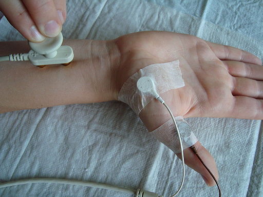
In the Nerve conduction study, assessment must be done for ABCDEFGH.
- Action potential Amplitude
- Measures the height of response in millivolts (mV) for motor nerve and microvolts (μV) for sensory nerve
- Indicates quantity of axons contributing to action potential
- Block of conduction
- CMAP on proximal stimulation is observed to be smaller than the CAMP on distal stimulation (reduced number of motor units have conducted the action potential over intervening segment of the nerve – feature of potentially recoverable neuropraxia)
- Conduction velocity
- Distance (between stimulating and recording electrode) divided by Duration
- Indicate quality of conduction along the axons
- Upper limb: 50 m/s (motor)
- Lower limb: 40 m/s (motor)
- Sensory conduction velocity: about 10 m/s faster
- Dispersion (Temporal dispersion)
- A reduction in the proximal CMAP compared to distal CMAP amplitude with the proximal CMAP duration increases by 20% or more
- Delay (Latency)
- Measures the time between onset of the stimulus and the response in milliseconds (ms)
- Indicate quality of conduction along the axons
- Upper limb: <3-4.5 ms (motor) and <2.3-3.5 ms (sensory)
- Lower limb: <6-6.5 ms (motor) and <2.6-2.9 ms (sensory)
- Echo – F wave
- The F wave is a kind of electrical echo in which the impulse travels up to the spine and then back down along the same fiber. It thereby gives a sense of the conduction along the entire length of a motor nerve.
- Uses supramaximal stimulation
- Absent in early proximal lesions (root or proximal injuries)
- Gastrocnemius contraction – H reflex
- The H reflex is basically an electrophysiologically recorded Achilles muscle stretch reflex. It is performed by stimulating the tibial nerve in the popliteal fossa. From there, the stimulus goes proximally through the reflex arc at that spinal segment, then distally from the anterior horn cell and the motor nerve. It can be recorded over the soleus or gastrocnemius muscles. The H reflex is most commonly used to evaluate for an S1 radiculopathy or to distinguish from an L5 radiculopathy.
- Uses submaximal stimulation
The fastest motor nerve conduction velocity (FMNCV) is reduced by approximately 1 m/s per °C temperature fall. The motor conduction slows by 0.4–1.7 m/s per decade after 20 years and the sensory by 2–4 m/s.
| Parameters | Axonal (Axon) loss | Demyelination |
|---|---|---|
| Sensory responses | Small or absent | Small or absent |
| Distal motor latency | Normal or slightly prolonged | Prolonged |
| CMAP amplitude | Small | Normal (reduced if conduction block or temporal dispersion) |
| Conduction block/temporal dispersion | Not present | Present |
| Motor conduction velocity | Normal or slightly reduced (<2/3rd) | Notably reduced (>2/3rd) |
| F waves minimum latency | Normal or slightly prolonged | Significantly prolonged |
Loss of Sensory Nerve Action Potential (SNAP) reflects a lesion distal to the Dorsal Root Ganglion (spinal foramen) like in plexus. Intact SNAP in a hypaesthetic limb suggests disease proximal to the Dorsal Root Ganglion (spinal foramen) like in a prolapsed disc.
Electromyography (EMG)
In the EMG, assessment must be done for activity during:
- Needle insertion (Insertional activity)
- Contraction of muscle (Muscle unit potential)
- Rest of muscle (Spontaneous activity)
- Abnormal activity during contraction (Interference pattern)
| EMG stage | Normal | Neurogenic lesion – UMNL | Myogenic lesion |
| Insertional activity | Normal (brief) | Normal – Increased | Normal – Increased, myotonic discharges |
| Spontaneous activity | Normal (silent) | Normal (silent) Fibrillation potentials Positive sharp waves (Muscles sometimes start having spontaneous activity on their own) | Normal (silent) Fibrillation potentials Positive sharp waves (Inflammation or muscle disease) |
| Motor unit potential | 0.5-1 mV amplitude; 5-10 ms duration; Normal recruitment | Normal to increased amplitude and duration Normal to decreased recruitment (Nerve is unable to connect many motor units for recruitment) | Small amplitude Early recruitment (insufficient force is generated by any given motor unit, and additional motor units are recruited earlier or more rapidly than expected) Myotonic discharges |
| Interference pattern | Full | Reduced | Full, low amplitude |


