Skip to content
Epomedicine

Mnemonics, Simplified Concepts & Thoughts

Epomedicine

Mnemonics, Simplified Concepts & Thoughts

Internal and External Jugular Veins with Mnemonics

Epomedicine, May 18, 2024

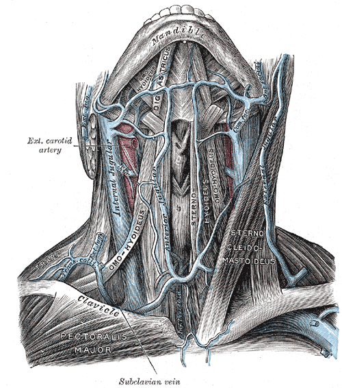
Jugular veins are the veins that take blood from the head to the heart via Superior Venacava (SVC).

jugular veins
Henry Vandyke Carter, Public domain, via Wikimedia Commons
Internal Jugular Vein (IJV)External Jugular Vein (EJV)
Formation (Drains from)Inferior petrosal sinus + Sigmoid sinus (At or just distal to Jugular foramen)Posterior div. of retromandibular vein + Posterior auricular vein (Within parotid gland at the angle of mandible)
CourseCarotid sheath (lateral-most)Subcutaneous anterolateral neck (superficial then posterior to sternocleidomastoid)
TributariesMnemonic: Medical Schools Let Fun People InMnemonic: PAST
1. Middle thyroid vein
2. Superior thyroid vein
3. Lingual vein
4. common Facial vein (facial vein + ant. branch of retromandibular vein)
5. Pharyngeal plexus
6. Inferior petrosal sinus
1. Posterior external jugular vein
2. Anterior jugular vein
3. Suprascapular vein
4. Transverse cervical vein
Termination (Drains to)Brachiocephalic vein (posterior to sternal end of clavicle deep to sternocleidomastoid)Subclavian vein
Drainage areaMajor venous return from Brain, Upper face and NeckScalp, Face and part of Scapular region
Clinical significanceJugular venous pulse and pressureJugular vein distension
  • Facebook
  • Twitter
PGMEE, MRCS, USMLE, MBBS, MD/MS AnatomyCardiovascular system

Post navigation

Previous post
Next post

Related Posts

PGMEE, MRCS, USMLE, MBBS, MD/MS

Coccyx Types

May 23, 2025May 23, 2025

Postacchini and Massobrio Classification (Nathan Modification) Can be classified into 6 types: Type I (slight curve): Curved slightly forward with apex pointing caudally (>50% incidence) Type II (forward curve): More prominent ventral curvature with coccyx apex pointing anteriorly (8-32% incidence) Type III (sharp angle): Acute anterior angulation of coccyx but…

Read More
PGMEE, MRCS, USMLE, MBBS, MD/MS midbrain cross-section

How to Draw Midbrain Cross-section ?

Jul 17, 2016Jul 17, 2016

The cross-section of midbrain can be compared to the “upside down striped face of a red-eyed demon“. Using this analogy of a demon face, lets assign the structures found on the cross-section of midbrain: Ear = Crus cerebri Medial – frontopontine fibers Middle – corticonuclear and corticospinal tract Lateral –…

Read More
PGMEE, MRCS, USMLE, MBBS, MD/MS protein-c-s mnemonic

Protein C and S pathway – Mnemonic

May 23, 2019May 23, 2019

Factor Va and VIIIa are different in coagulation cascade. They are the co-factors for factor Xa and IXa respectively. Another mnemonic: protein C Cuts Coagulation by Cutting Cofactors (Va and VIIIa) Protein S is cofactor for protein S. Protein C and S pathway using the mnemonic: Time To: Thrombin (factor…

Read More

Leave a Reply Cancel reply

Your email address will not be published. Required fields are marked *

This site uses Akismet to reduce spam. Learn how your comment data is processed.

Epomedicine. Internal and External Jugular Veins with Mnemonics [Internet]. Epomedicine; 2024 May 18 [cited 2025 Dec 15]. Available from: https://epomedicine.com/medical-students/internal-and-external-jugular-veins-with-mnemonics/.

Pre-clinical (Basic Sciences)

Anatomy

Biochemistry

Community medicine (PSM)

Embryology

Microbiology

Pathology

Pharmacology

Physiology

Clinical Sciences

Anesthesia

Dermatology

Emergency medicine

Forensic

Internal medicine

Gynecology & Obstetrics

Oncology

Ophthalmology

Orthopedics

Otorhinolaryngology (ENT)

Pediatrics

Psychiatry

Radiology

Surgery

RSS Ask Epomedicine

  • What to study for Clinical examination in Orthopedics?
  • What is the mechanism of AVNRT?

Epomedicine weekly

  • About Epomedicine
  • Contact Us
  • Author Guidelines
  • Submit Article
  • Editorial Board
  • USMLE
  • MRCS
  • Thesis
©2025 Epomedicine . All rights reserved.