Skip to content
Epomedicine

Mnemonics, Simplified Concepts & Thoughts

Epomedicine

Mnemonics, Simplified Concepts & Thoughts

painless loss of vision

Vision loss – General Appoach

Epomedicine, Aug 15, 2015Aug 17, 2015

Diagnosis of the etiology of vision loss requires a step-wise systematic approach. The vision loss may be sudden or gradual, painful or painless, unilateral or bilateral, etc. One needs to take a detailed ocular history and examination.

Step 1: Sudden or gradual vision loss?

a. Sudden: vascular occlusion (e.g., AION, CRAO, CRVO) or bleeding (e.g., vitreous hemorrhage, “wet” macular degeneration).

b. Gradual: degenerations or depositions (e.g., cataract, macular dystrophies or “dry” macular degeneration, corneal dystrophies).

Step 2: Associated with pain?

  • Associated pain is a common finding in anterior ocular processes (e.g., keratitis, anterior uveitis)
  • Other painful conditions: orbital disease, optic neuritis, and giant cell arteritis

painless vision loss

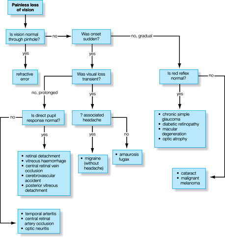
Step 3: Transient or persistent vision loss?

a. Transient: temporary/subcritical vascular insufficiency (e.g., giant cell arteritis, amaurosis fugax, vertebrobasilar
artery insufficiency) or papilledema

b. Persistent: structural or irreversible damage (e.g., vitreous hemorrhage, macular degeneration).

Step 4: Unilateral or Bilateral vision loss?

a. Unilateral: a local cause or lesion anterior to optic chiasma

b. Bilateral: a more widespread or systemic process or lesion posterior to optic chiasma

Step 5: Blurred, dimmed or distorted vision (What)?

a. Blurring or dimming: pathology anywhere in the visual pathway from cornea to cortex; common problems include refractive error, cataract, and macular disease.

b. Distortion: macular pathology, high refractive error (high ametropia/astigmatism) or other ocular disease.

Step 6: Where is the vision loss?

a. Superior or inferior hemispheric field loss (Altitudinal): corresponding inferior or superior vascular event involving the retina (e.g., retinal vein occlusion) or optic disc (e.g., segmental AION)

b. Peripheral field loss: retinal detachment (usually rapidly evolving from far periphery), optic nerve disease, chiasmal compression (typically bitemporal loss), or cortical pathology (homonymous hemianopic defects)

c. Central blurring of vision: diseases of the macula (positive scotoma: a “seen” spot) or optic nerve (negative scotoma: an unseen defect).

Step 7: When is the vision loss ?

Glare: from headlights or bright sunlight due to posterior subcapsular lens opacities

Further investigations is based upon the disease suspected from history and ophthalmic examination.

Adapted from Oxford Handbook of Ophthalmology

6 shares
  • Facebook6
  • Twitter
PGMEE, MRCS, USMLE, MBBS, MD/MS Ophthalmology

Post navigation

Previous post
Next post

Related Posts

PGMEE, MRCS, USMLE, MBBS, MD/MS hematopoiesis

Hematopoietic growth factors list with mnemonics

May 23, 2019May 23, 2019

a. SCF (Steel factor): Multipontent stem cells b. IL-3: Myeloid stem cells (“3” turned down looks like “m“) c. IL-7: Lymphoid stem cell (“L” turned up looks like “7“) d. GM-CSF (Granulocyte macrophage Colony stimulating factor): Myeloid lineage (Erythrocytes, Thrombocytes, Granulocytes and Monocytes) Pharmaceutical: Sargramostim e. G-CSF: Granulocytes (Neutrophil, Eosinophil,…

Read More
PGMEE, MRCS, USMLE, MBBS, MD/MS

Synovial Fluid Analysis – Made Easy

Jun 13, 2020Jun 13, 2020

What investigations to order for synovial fluid sample? Mnemonic: 5 Cs Chemistry (pH, LDH, glucose, protein) Cell counts Cytology Culture Crystals Interpretation of synovial fluid analysis Gross Normal Non-inflammatory Inflammatory Septic Crystal Hemorrhagic Volume (ml) <3.5 >3.5 >3.5 >3.5 >3.5 >3.5 Viscosity High High Low Variable Variable Variable Color Colorless…

Read More
PGMEE, MRCS, USMLE, MBBS, MD/MS spinal tracts

Spinal Tracts Simplified

Jul 25, 2016May 16, 2024

Organization of Ascending and Descending Tracts in Spinal Cord A. 2 Posterior Tracts: The fibers of these tracts cross to the opposite side at the level of medulla: B. 2 Lateral Tracts: The fibers of these tracts remain on ipsilateral side: C. 2 Anterior Tracts: The fibers of these tracts…

Read More

Leave a Reply Cancel reply

Your email address will not be published. Required fields are marked *

This site uses Akismet to reduce spam. Learn how your comment data is processed.

Pre-clinical (Basic Sciences)

Anatomy

Biochemistry

Community medicine (PSM)

Embryology

Microbiology

Pathology

Pharmacology

Physiology

Clinical Sciences

Anesthesia

Dermatology

Emergency medicine

Forensic

Internal medicine

Gynecology & Obstetrics

Oncology

Ophthalmology

Orthopedics

Otorhinolaryngology (ENT)

Pediatrics

Psychiatry

Radiology

Surgery

RSS Ask Epomedicine

  • What to study for Clinical examination in Orthopedics?
  • What is the mechanism of AVNRT?

Epomedicine weekly

  • About Epomedicine
  • Contact Us
  • Author Guidelines
  • Submit Article
  • Editorial Board
  • USMLE
  • MRCS
  • Thesis
©2026 Epomedicine | WordPress Theme by SuperbThemes