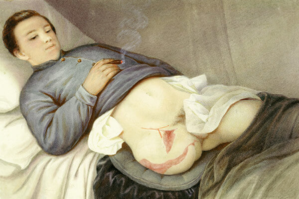
Indications
Mnemonic: three Ds
1. Dead (or dying): Peripheral vascular disease (most common indication for amputation), Severe trauma (leading indication for amputation in younger patients), Burns, Frostbite
- Most significant predictor of amputation in diabetics is peripheral neuropathy, as measured by insensitivity to the Semmes-Weinstein 5.07 monofilament
2. Dangerous (or deadly): Malignant tumors, Potentially lethal sepsis, Crush injury
3. Damned nuisance: Gross deformity, Recurrent sepsis, Sever loss of function
Increase in Energy Expenditure above Baseline
Energy required for walking is inversely proportional to the length of the remaining limb. Traumatic amputees are generally younger and vascular amputees are generally older. As the level of amputation moves proximally, the walking speed decreases, and the oxygen consumption increases.
1. Syme’s (ankle): 15%
2. Transtibial (BKA): 25% in traumatic (10% long transtibial, 40% short transtibial), 40% in vascular
3. Transfemoral (AKA): 65% in traumatic, 100% in vascular
4. Hindquarter: 100%
Walking velocity
Slower rates for amputees seem to be a compensatory mechanism to conserve energy per unit time.
a. Vascular amputees:
- Syme: 66%
- Transtibial: 59%
- Transfemoral: 44%
b. Traumatic amputees:
- Transtibial: 87%
- Transfemoral: 63%
Level of amputation
Requires an understanding of the tradeoffs between increased function with a more distal level of amputation and a decreased complication with a more proximal level of amputation.
1. Malignant tumor, life threatening infection or irreparable damaged body part: Proximal to lesion in healthy tissue
2. Peripheral vascular disease: Predictors of wound healing are –
- Ankle brachial index (ABI) >0.45
- Doppler ultrasonography pressure >70 mmHg
- Ischemic index (II) >0.5 (ABI/Systolic pressure at brachial artery)
- Transcutaneous oxygen measurement >30-40 mmHg
- Toe pressure >40 mmHg
- Hair on leg or dorsum of foot
- TLC >1500/cu.mm
Thermal gradient from proximal to distal and skin trophic changes: clinical sign of poor vascular supply to the soft tissue envelope
3. Peripheral ischemia secondary to frostbite, vasoconstrictor administration for hypotension and cryoglobulinemia:
- Allow time for completion of tissue demarcation and to keep the necrotic areas dry (allow autoamputations of the necrotic portions)
- Technetium-99 bone scanning helps to demarcate viable from non-viable tissues
- May take 2-6 months
4. Contracted knee despite intensive physiotherapy: Knee disarticulation instead of BKA
5. Non-ambulator patients: Select level that will ensure best chance of healing
6. Gangrenous changes of heel pad: BKA
General principles of technique
1. Tourniquet and Exsanguination:
- Contraindication for tourniquet: Arterial insufficiency
- Contraindication for exsanguination: Infection and Malignancy
2. Skin flaps:
- Length: Combined length = 1.5 X width or diameter of limb (d)
- Diameter (d) = Circumference/3.14
- Equal flaps = 3/4 X d Anterior + 3/4 X d Posterior flap = 1.5 X d
- Unequal flaps = 1/2 X d Anterior + 1 X d Posterior flap = 1.5 X d
- Flap length selection:
- Upper limb: Equal anterior and posterior flap
- Trans-femoral: Equal anterior and posterior flap
- Trans-tibial: Long posterior flap
- Keep thick (avoid unnecessary dissection)
- Should not be adherent to underlying bone
- Avoid large “dog ears”
3. Muscle flaps:
- Length: Sectioned 5 cm distal to level of intended bone resection
- Stabilization:
- Myoplasty: Suturing muscle to periosteum or fascia of opposing musculature
- Myodesis: Suturing muscle or tendon to bone through drill holes
- Advantages: Provide a stronger insertion, help maximize strength, minimize atrophy (transected muscles atrophy 40-60% in 2 years if not fixed securely), counterbalance antagonist muscles (prevent contractures and maximize residual limb function) and provides firm stump
- Contraindication: Severe ischemia (may further compromise already marginal blood supply)
- Above knee amputation in non-ischemic limb: Adductor magnus and Hamstring myodesis
- Above elbow amputation in non-ischemic limb: Triceps tenodesis
- Osteomyodesis: Similar to myodesis but the periosteum is stripped (enables bone growth in that area)
Advantages of myoplasty/myodesis:
a. Good stump shape
b. Muscles insulate cut nerve endings and bone from prosthesis
c. Muscles originating proximal to joint produce better stump mobility and increase leverage
d. Muscles not acting on joint contract isometrically and assist in venous return
e. Prevent retraction and painful muscle contractions
f. Prevents phantom pain
4. Blood vessels and Hemostasis:
- Isolated and individually ligated
- Large blood vessels doubly ligated
5. Nerves:
- Divided under tension (avoid strong tension) proximal to bone with a sharp blade cut to avoid neuroma on weightbearing area
- Techniques to prevent neuroma: end loop anastomosis, perineural closure, silastic capping, sealing epineural tube with butyl-cyanoacrylate, cauterization, or burying nerve ends in muscle/bone
- Large nerves like sciatic nerve often contain relatively large vessels and should be ligated
6. Bone:
- Sawn across at the proposed level:
- Upper limb
- Above elbow: 20 cm from acromion (attempt to preserve 5 to 7 cm of the proximal humerus)
- Below elbow: 18 cm from olecranon (attempt to preserve at-least 5 cm ulna in proximal amputations)
- Lower limb
- Above knee: 12 cm from joint line
- Trans-femoral residual limb length:
- Short: 0-35% of femur remains
- Standard: 35-60% of femur remains
- Long: >60% of femur remains
- Trans-femoral residual limb length:
- Below knee: 14 cm from joint line
- Non-ischemic limb: 12.5-17.5 cm below joint line depending on body height (2.5 cm bone length for each 30 cm body height)
- Ischemic limb: Higher level (8.8-12.5 cm below joint line)
- Bevel tibia on anterior and medial aspect
- Fibula: Cut 3 cm proximal to tibia
- Ertl technique: Creation of a tibiofibular bone bridge (fibular strut autograft is secured with heavy suture and tibial periosteal sleeve is sewn around strut distally) provides a stable, broad tibiofibular articulation that may be capable of some distal weight bearing
- Trans-tibial residual limb length:
- Short: <20% of limb below the knee remains
- Standard: 20-50% of limb below the knee remains
- Long: 50-90% of limb below the knee remains
- Above knee: 12 cm from joint line
- Upper limb
- Avoid excessive periosteal stripping (to prevent ring sequestrum or bony overgrowth)
- Rasp the bone to form smooth contour
7. Wound closure and dressing:
- Tourniquet deflated and hemostasis reassured before closure
- Drain preferable for 48-72 hours
- Dressing:
- Conventional soft dressing (using elastic bandage in figure of 8 fashion): Indicated when amputation wound requires frequent observation e.g. infection
- Rigid dressing: Total contact Plaster of Paris cast
- Types:
- Rigid dressing without immediate postop prosthesis (IPOP)
- Rigid dressing with immediate postop prosthesis (IPOP) – allows weight bearing within 24 hours
- Removable rigid dressing (RRD)
- Advantages:
- Prevent edema at surgical site
- Protect wound from bed trauma
- Enhance wound healing
- Early maturation of stump
- Decrease postoperative pain
- Allow early mobilization from bed
- Prevent knee flexion contractures
- Types:
Rehabilitation
1. Acute postsurgical (POD 1-4): Wound healing, pain control, stump positioning education, precautions to prevent flexion and abduction contractures (firm mattress, lying prone QID X 15 minutes, promoting knee extension while resting) proximal body motion, emotional support, phantom limb discussion
2. Preprosthetic rehabilitation (POD 4 – 3 weeks):
- Muscle setting and joint mobilization exercises
- Start prosthetic gait training with parallelbars and progress to walkers and cane (avoid crutch for due to risk of developing poor gait mechanics)
- Remove rigid dressing at 1 week (if wound complications not suspected) and change weekly until volume appears unchanged from previous week
3. Prosthetic training (after weeks 3-4):
- Suture or staple removal
- Temporary prosthesis fitting
- Continued physiotherapy
- Definitive prosthesis is usually created at 3 to 6 months
Complications
A. Early:
1. Hematoma
- Prevented by meticulous hemostasis and rigid dressing
- May delay wound healing and act as nidus for infection
- Treat with compressive dressing +/- evacuation
2. Infection
- More common in diabetic, ischemic limbs
- Risk of gas gangrene with high above knee amputations (from perineum)
- Deep wound infections: Treat with immediate debridement, irrigation and open wound management
- Smith and Burgess method: Central 1/3 of wound is closed, and remainder of wound is packed (allows for continued open wound management while maintaining adequate flaps for distal bone coverage)
3. Wound necrosis
- Nutritional assessment (Serum albumin, TLC) and supplementation, Smoking cessation (if present)
- Necrosis of skin edge <1 cm: Treat conservatively with open wound management
- Discontinue prosthesis until wound healed
- Severe necrosis with loss of bone coverage: Wedge resection (incorporating the full diameter of the stump would allow for reformation of the hemisphere while minimizing local pressures)
B. Late complications
1. Contractures
- Prevented by proper stump positioning, gentle passive stretching, exercises and increased ambulation
- Prosthetic modification may be necessary
- May need wedging casts or surgical release of contracted structures
2. Chronic pain
- Residual limb pain:
- Poorly fitting prosthesis: Socket modification
- Painful neuroma: Socket modification → failed → Neuroma excision or proximal neurectomy (excising 3 cm of nerve above the neuroma)
- Other causes: Osteoarthritis of hip or knee, Referred pain from back
- Phantom limb sensations:
- Common and not bothersome usually
- Telescoping: Phantom limb gradually shortens to residual limb (during 1st year)
- Phantom limb pain:
- Less common
- More common with proximal amputations
- May be beneficial: massage, ice, heat, increased prosthetic use, relaxation training, biofeedback, sympathetic blockade, oral medications, local nerve blocks, epidural blocks, ultrasound, transcutaneous electrical nerve stimulation, and placement of a dorsal column stimulator
7. Dermatological problems
- Eczema
- Bacterial folliculitis
- Epidermoid cysts
- Verrucous hyperplasia
- caused by proximal constriction that prevents the stump from fully seating in the prosthesis (“choking”) causes distal stump edema followed by thickening of the skin, fissuring, ulceration, and possibly subsequent infection
- socket modification is mandatory
