Skip to content
Epomedicine

Mnemonics, Simplified Concepts & Thoughts

Epomedicine

Mnemonics, Simplified Concepts & Thoughts

eye schematic

Embryology of Eye : Mnemonic

Epomedicine, Mar 3, 2019Mar 3, 2019

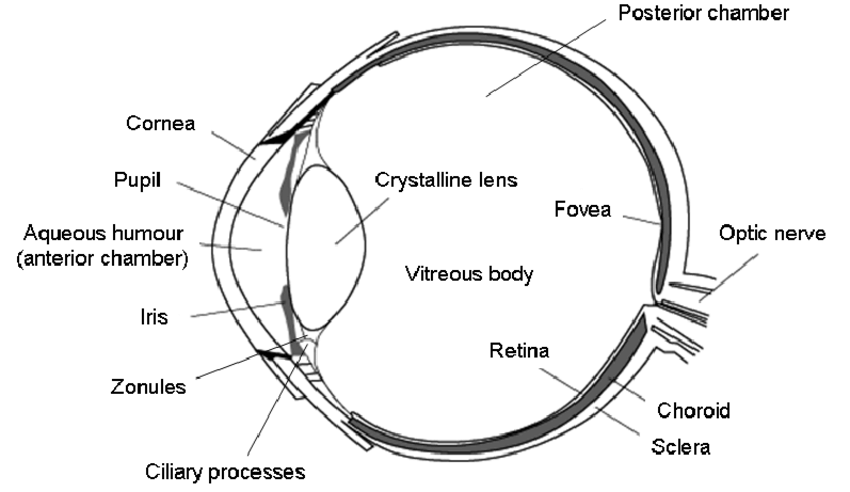
Surface ectoderm derivatives

Mnemonic: LEVeL

  1. Lens
  2. Epithelium
    • All ocular adnexa including mebomian gland and glands of Zeis and Moll
    • Skin
    • Cornea
    • Conjunctiva
  3. Vitreous (portion)
  4. Lacrimal glands and drainage system

Neuro-ectoderm (Optic vesicle and cup) derivatives

Mnemonic: MORE

  1. Muscles of pupil (constrictor and dilator pupillae)
  2. Optic nerve
  3. Retinal pigment epithelium
  4. Epithelium of iris and ciliary body

Mesoderm derivatives

Mnemonic: MESO

  1. Muscles (Extraocular)
  2. Endothelium of eye and orbital blood vessels
  3. Sclera (temporal portion)
  4. Others (Vitreous)

All other structures are derived from Neural crest cells.

55 shares
  • Facebook53
  • Twitter
PGMEE, MRCS, USMLE, MBBS, MD/MS AnatomyEmbryologyOphthalmology

Post navigation

Previous post
Next post

Related Posts

PGMEE, MRCS, USMLE, MBBS, MD/MS

Popliteal Fossa : Mnemonics

May 13, 2023Aug 9, 2023

Mnemonic: Serve And Volley Next Ball Think of popliteal fossa as a diamond-shaped space. The floor is formed by Popliteus muscle. Membranosus = Medial (The 2 “Semis” go together – hence medial border is formed by semimembranosus, semitendinosus and medial head of gastrocnemius) Therefore, biceps femoris and lateral head of…

Read More
PGMEE, MRCS, USMLE, MBBS, MD/MS

Compartments, Muscles and Fasciotomy of the leg

Jul 27, 2022Jul 27, 2022

Muscle Origin Insertion Action Innervation Anterior compartment 1. Tibialis anterior (TA) Superior 2/3 lateral surface of tibia Medial cuneiform, 1st metatarsal Dorsiflexion, foot inversion Deep peroneal nerve (L5) 2. Extensor digitorum longus (EDL) Superior 2/3 of fibula and interosseous membrane Middle and distal phalanx, lateral 4 toes Dorsiflexion, toe extension…

Read More
PGMEE, MRCS, USMLE, MBBS, MD/MS parinaud syndrome

Parinaud Syndrome – Anatomical Basis

May 19, 2019May 19, 2019

Synonyms: Dorsal midbrain sydnrome, Pretectal syndrome Main components of Parinaud syndrome: Vertical gaze palsy Convergence-retraction nystagmus Pupillary light-near dissociation Lid retraction (Collier’s sign) Anatomical basis of Parinaud syndrome: Site of lesion: Dorsal midbrain Close to cerebral aqueduct and pineal gland – hydrocephalus and pinealoma Arterial supply – posterior cerebral artery…

Read More

Leave a Reply Cancel reply

Your email address will not be published. Required fields are marked *

This site uses Akismet to reduce spam. Learn how your comment data is processed.

Pre-clinical (Basic Sciences)

Anatomy

Biochemistry

Community medicine (PSM)

Embryology

Microbiology

Pathology

Pharmacology

Physiology

Clinical Sciences

Anesthesia

Dermatology

Emergency medicine

Forensic

Internal medicine

Gynecology & Obstetrics

Oncology

Ophthalmology

Orthopedics

Otorhinolaryngology (ENT)

Pediatrics

Psychiatry

Radiology

Surgery

RSS Ask Epomedicine

  • What to study for Clinical examination in Orthopedics?
  • What is the mechanism of AVNRT?

Epomedicine weekly

  • About Epomedicine
  • Contact Us
  • Author Guidelines
  • Submit Article
  • Editorial Board
  • USMLE
  • MRCS
  • Thesis
©2026 Epomedicine | WordPress Theme by SuperbThemes