Skip to content
Epomedicine

Mnemonics, Simplified Concepts & Thoughts

Epomedicine

Mnemonics, Simplified Concepts & Thoughts

Cervical Vertebrae Anatomy

Epomedicine, May 4, 2024May 4, 2024

Osteology

cervical vertebrae
“#atlas #axis #cervical #thoracic #spine #vertebra #bones #perth #tafe #work #anatomy” by fitnessferg is licensed under CC BY-NC-SA 2.0.
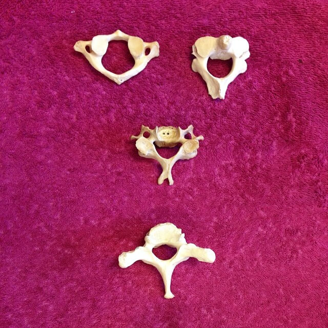
C1 (top left), C2 (top right), Typical vertebra (middle), C7 (down)
VertebraBodySpinous processTransverse process
Typical (C3-6)Relatively small; oval; uncinate process superiorlyBifidHas foramen transversarium, Has anterior and posterior tubercle separated by groove for spinal nerve (large anterior tubercle in C6 called Carotid/Chassaignac tubercle)
Atlas (C1)None; Ring (2 lateral masses connected by anterior & posterior arches)None (has posterior tubercle)Has foramen transversarium; No tubercles
Axis (C2)Has dens (odontoid process)BifidHas foramen transversarium; Has 1 tubercle (posterior)
C7Larger; uncinate process superiorlyUnifid (longest – vertebra prominens)Has foramen transversarium (may be small or absent; transmits vein only/doesn’t transmit artery); Has 1 tubercle (posterior)

Joints or Articulations

JointArticulationsJoint typeFunctions
Atlanto-occipital (X2)Superior facets of lateral mass (C1) + Occipital condyles (Cranium base)Condyloid“Yes” nodding (flexion-extension)
Atlanto-axial (X3)Lateral = Inferior facets of lateral mass (C1) + Superior facets of C2
Medial = Dens (C2) + Facet for dens (C1)
Lateral (Plane)
Medial (Pivot)
“No” movement (rotation)
OthersBetween vertebral arches (Zygoapophyseal/Facet X 2) = Inferior facets of superior vertebra + Superior facets of inferior vertebra
Vertebral bodies (Intervertebral X 1) = 2 vertebral bodies articulated by a “disc”
Vertebral bodies (Uncovertebral/Luschka X 2) = Uncinate process of inferior vertebra + Inferolateral aspect of superior vertebra
Zygoapophyseal (synovial)
Intervertebral (symphysis/fibrocartilaginous)
Uncovertebral (pseudojoints)
Flexion, Extension, Rotation

Ligaments

  1. Supraspinous ligament (Liagametum nuchae): Runs between tips of 2 spinous process
  2. Interspinous ligament: Between 2 spinous processes
  3. Ligamentum flavum: Connects the laminae of adjacent vertebrae
  4. Intertransverse ligaments: Run between adjacent transverse processes
  5. Anterior longitudinal ligament: Between 2 vertebral body (not attached to disc) from sacrum to C2
    • C1-C2: Anterior atlantoaxial membrane
    • C1-Oc: Anterior atlanto-occipital membrane
  6. Posterior longitudinal ligament: Between 2 vertebral body (firmly attached to disc) from sacrum to C2
    • C2-Foramen magnum: Tectorial membrane
  7. Apical ligament: Connects apex of dens of C2 to foramen magnum (anterior aspect)
  8. Alar ligaments: Connects dens of C2 to foramen magnum (lateral aspect)
  9. Cruciate liagments: Vertical ligaments (Dens to occipital bone and body of C2) + Transverse ligaments (Dens to arch of C1)
  • Facebook
  • Twitter
PGMEE, MRCS, USMLE, MBBS, MD/MS AnatomyMusculoskeletal system

Post navigation

Previous post
Next post

Related Posts

PGMEE, MRCS, USMLE, MBBS, MD/MS Child Pugh Score

Hepatorenal syndrome (HRS) – Quick revision

Apr 18, 2017

New Criteria for HRS 1. Cirrhosis with ascites 2. Serum creatinine >1.5mg/dl 3. No sustained improvement in renal function after 2 days of diuretic withdrawl (if on diuretics) and volume expansion with albumin infusion at 1 gm/kg/day upto a maximum of 100 gm/day. 4. No evidence of shock 5. No…

Read More
PGMEE, MRCS, USMLE, MBBS, MD/MS Advanced glaucomatous changes diagram

Glaucomatous Optic Disc Changes Made Simple

May 3, 2014May 5, 2014

Optic disc changes in Glaucoma is one of the most frequently asked topics in Ophthalmology. However, students are often found to have difficulty in understanding and remembering them. Hence, I came up with an idea to illustrate them with schematic diagrams and mnemonics to simplify the topic. The diagrams are…

Read More
PGMEE, MRCS, USMLE, MBBS, MD/MS granuloma morphology pathophysiology

Morphology of Granuloma

Jan 19, 2017Jan 31, 2017

Concentric layers of Granuloma There are 4 concentric layers in a granuloma, however the clear distinction is difficult in reality due to overlapping. From inside to out: 1. Necrosis Caseating necrosis: Tuberculosis, Leprosy Coagulative necrosis: Buruli ulcer (M.ulcerans), Gumma containing central blood vessels (Syphilis) Fibrinoid necrosis: Aschoff bodies (Rheumatic granuloma),…

Read More

Leave a Reply Cancel reply

Your email address will not be published. Required fields are marked *

This site uses Akismet to reduce spam. Learn how your comment data is processed.

Epomedicine. Cervical Vertebrae Anatomy [Internet]. Epomedicine; 2024 May 4 [cited 2026 May 21]. Available from: https://epomedicine.com/medical-students/cervical-vertebrae-anatomy/.

Pre-clinical (Basic Sciences)

Anatomy

Biochemistry

Community medicine (PSM)

Embryology

Microbiology

Pathology

Pharmacology

Physiology

Clinical Sciences

Anesthesia

Dermatology

Emergency medicine

Forensic

Internal medicine

Gynecology & Obstetrics

Oncology

Ophthalmology

Orthopedics

Otorhinolaryngology (ENT)

Pediatrics

Psychiatry

Radiology

Surgery

RSS Ask Epomedicine

  • What to study for Clinical examination in Orthopedics?
  • What is the mechanism of AVNRT?

Epomedicine weekly

  • About Epomedicine
  • Contact Us
  • Author Guidelines
  • Submit Article
  • Editorial Board
  • USMLE
  • MRCS
  • Thesis
©2026 Epomedicine | WordPress Theme by SuperbThemes