Synonyms: Calot triangle, Cystohepatic triangle
Boundaries:
Cysto-hepatic triangle (Budde-Rocko triangle):
- Cystic artery
- Cystic duct
- Common hepatic duct
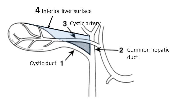
Calot’s triangle as described in modern surgery:
- Inferior: Cystic duct
- Medial: Common hepatic duct
- Above: Inferior liver surface (right lobe)
Mnemonic: 3 C
1. Cystic duct
2. Common hepatic duct
3. Cystic artery
Contents:
- Cystic artery
- Right branch of hepatic artery
- Occassionally accessory hepatic ducts and arteries
- Bile duct
- Cystic lymph node of Lund (Mascagni’s lymph node or Calot’s node)
- Connective tissue
- Lymphatics
Importance:
The cystic artery and the duct have to be clearly defined to obtain the ‘critical view of safety’. These structures are exposed by careful dissection of the fibrofatty tissue within the Calot’s triangle. Once the fibrofatty tissue is cleared, the cystic artery and the cystic duct are conclusively identified as the only two structures passing into the gallbladder and base of the liver bed is exposed by detaching the lowest part of the gallbladder from the liver. It is not necessary to see the common bile duct (CBD). This is the critical view of safety and once this is obtained, the cystic duct and artery can be safely clipped. If these structures are clearly seen, then even in the presence of abnormal anatomy an iatrogenic injury should be avoided.
Rouviere’s sulcus, a naturally occurring cleft in the right lobe, anterior to Segment 1, occurs in over 80% of normal livers. It is a useful, but often ignored, anatomical landmark for beginning dissection of Calot’s triangle, and also for confirming its location.
Steps of Laparoscopic cholecystectomy:
- Carboperitoneum
- Critical view of safety dissection
- Cystic duct ligation/division
- Cystic artery ligation/division
References:
- Rural Surgery: Challenges and Solutions for the Rural Surgeon edited by Matthias Wichmann, David C. Borgstrom, Nadine R. Caron, Guy Maddern
- Lockhart S, Singh-Ranger G. Rouviere’s sulcus-Aspects of incorporating this valuable sign for laparoscopic cholecystectomy. Asian J Surg. 2018 Jan;41(1):1-3. doi: 10.1016/j.asjsur.2016.07.012. Epub 2016 Sep 16. Review. PubMed PMID: 27647607.
- Laparoscopic Surgery of the Abdomen edited by Bruce MacFadyen