Skip to content
Epomedicine

Mnemonics, Simplified Concepts & Thoughts

Epomedicine

Mnemonics, Simplified Concepts & Thoughts

Applied anatomy of Anatomical Snuff Box

Epomedicine, Dec 17, 2013

Synonyms: Radial fossa, Foveola radialis

Note: Depression in the humerus which receives the head of radius is also named as Radial fossa

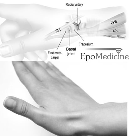
Definition: It is a triangular shaped depression in the radial or lateral aspect of the dorsum of the hand which is seen when the thumb is extended fully. This hollow was used to snuff powdered tobacco and hence named as Anatomical snuff box.

Boundaries and Borders:

  1. Medial border: Extensor Pollicis Longus (EPL)
  2. Lateral border: Extensor Pollicis Brevis (EPB) and Abductor Pollicis Longus (APL)
  3. Proximal border: Styloid process of radius
  4. Distal border: Base of 1st metacarpal
  5. Floor: Scaphoid and Trapezium bones
  6. Roof: Skin

Radial fossa

Contents:

  1. Radial artery
  2. Radial nerve: Dorsal cutaneous branch of radial nerve
  3. Cephalic vein

Note: Some books regard scaphoid and trapezium as contents of the snuff box.

Clinical Applications:

1. Differential diagnosis of Anatomical snuff box pain or tenderness:

  • DeQuervain’s tenosynovitis: Inflammation of the 1st extensor compartment of the wrist i.e. EPB and APL
  • Scaphoid fracture
  • Fracture of 1st metacarpal or distal radius
  • Scapholunate dislocation
  • Extensor carpi radialis or Flexor carpi radialis strain
  • Arthritis of Carpo-metacarpal or Radio-carpal joint
  • Cheiralgia paresthetica (handcuff neuropathy): compression or trauma of superficial branch of radial nerve
  • C6 cervical radiculopathy

2. Finkelstein’s maneuver (for DeQuervain’s tenosynovitis): Making a fist over thumb and ulnar deviation of wrist stretches the tendons of 1st extensor compartment causing significant pain.

Scaphoid tests
A. Finkelstein test
B. Scaphoid shift or Watson test

3. Signs of Scaphoid fracture:

  • Anatomical snuff box tenderness on palpation
  • Scaphoid tubercle tenderness (Location: extensor carpi radialis at palmar crease)
  • Scaphoid compression test (thumb is compressed against scaphoid)

4. Scaphoid shift or Watson test (Press scaphoid tubercle with thumb while moving wrist from ulnar to radial deviation): Pain or clunk suggests scapho-lunate instability

5. Why scaphoid is vulnerable to avascular necrosis?

  • 80% scaphoid surface is cartilage, leaving a small area for arterial blood supply to enter the bone.
  • Major blood supply is from dorsal carpal branch of radial artery which runs distal to proximal.
  • Facebook
  • Twitter
PGMEE, MRCS, USMLE, MBBS, MD/MS AnatomyMusculoskeletal systemOrthopedics

Post navigation

Previous post
Next post

Related Posts

PGMEE, MRCS, USMLE, MBBS, MD/MS CSF circulation

CSF Circulation Made Simple

Jul 17, 2016Jun 15, 2018

Cerebrospinal Fluid (CSF) Production and Absorption CSF is produced by the choroid plexus that lines the ventricles. Choroid plexus = Infoldings of blood vessels of piamater + Modified ciliated ependymal cells Tight junctions of the choroid plexus cells form Blood-CSF barrier. CSF is reabsorbed by arachnoid granulations to enter dural…

Read More
PGMEE, MRCS, USMLE, MBBS, MD/MS lymphatic drainage neck nodes level

Triangles and Lymph node levels of Neck

May 13, 2014Apr 29, 2024

There are 2 major triangles in the neck, containing other smaller triangles. 1. Anterior triangle: Midline of neck – Mandible – Anterior border of SCM Posterior triangle: Posterior border of SCM – Trapezius – Clavicle (level V); 2 triangles inside separated by inferior belly of omohyoid Surgically, cervical lymph nodes…

Read More
PGMEE, MRCS, USMLE, MBBS, MD/MS eye schematic

Embryology of Eye : Mnemonic

Mar 3, 2019Mar 3, 2019

Surface ectoderm derivatives Mnemonic: LEVeL Lens Epithelium All ocular adnexa including mebomian gland and glands of Zeis and Moll Skin Cornea Conjunctiva Vitreous (portion) Lacrimal glands and drainage system Neuro-ectoderm (Optic vesicle and cup) derivatives Mnemonic: MORE Muscles of pupil (constrictor and dilator pupillae) Optic nerve Retinal pigment epithelium Epithelium…

Read More

Leave a Reply Cancel reply

Your email address will not be published. Required fields are marked *

This site uses Akismet to reduce spam. Learn how your comment data is processed.

Epomedicine. Applied anatomy of Anatomical Snuff Box [Internet]. Epomedicine; 2013 Dec 17 [cited 2026 May 20]. Available from: https://epomedicine.com/medical-students/applied-anatomical-snuff-box/.

Pre-clinical (Basic Sciences)

Anatomy

Biochemistry

Community medicine (PSM)

Embryology

Microbiology

Pathology

Pharmacology

Physiology

Clinical Sciences

Anesthesia

Dermatology

Emergency medicine

Forensic

Internal medicine

Gynecology & Obstetrics

Oncology

Ophthalmology

Orthopedics

Otorhinolaryngology (ENT)

Pediatrics

Psychiatry

Radiology

Surgery

RSS Ask Epomedicine

  • What to study for Clinical examination in Orthopedics?
  • What is the mechanism of AVNRT?

Epomedicine weekly

  • About Epomedicine
  • Contact Us
  • Author Guidelines
  • Submit Article
  • Editorial Board
  • USMLE
  • MRCS
  • Thesis
©2026 Epomedicine | WordPress Theme by SuperbThemes