Skip to content
Epomedicine

Mnemonics, Simplified Concepts & Thoughts

Epomedicine

Mnemonics, Simplified Concepts & Thoughts

Anatomy of Physis (Growth Plate)

Epomedicine, Jul 30, 2024Jul 30, 2024

Periphery of physis:

The physis is connected to the epiphysis and metaphysis peripherally via:

  1. Groove of Ranvier (supplies chondrocytes to the periphery for latitudinal or appositional growth)
  2. Perichondral ring of LaCroix (strong fibrous tissue that anchors physis to metaphysis)

Blood supply of physis:

There are three sources of blood supply to the physis: the epiphyseal, metaphyseal, and perichondral circulations. 
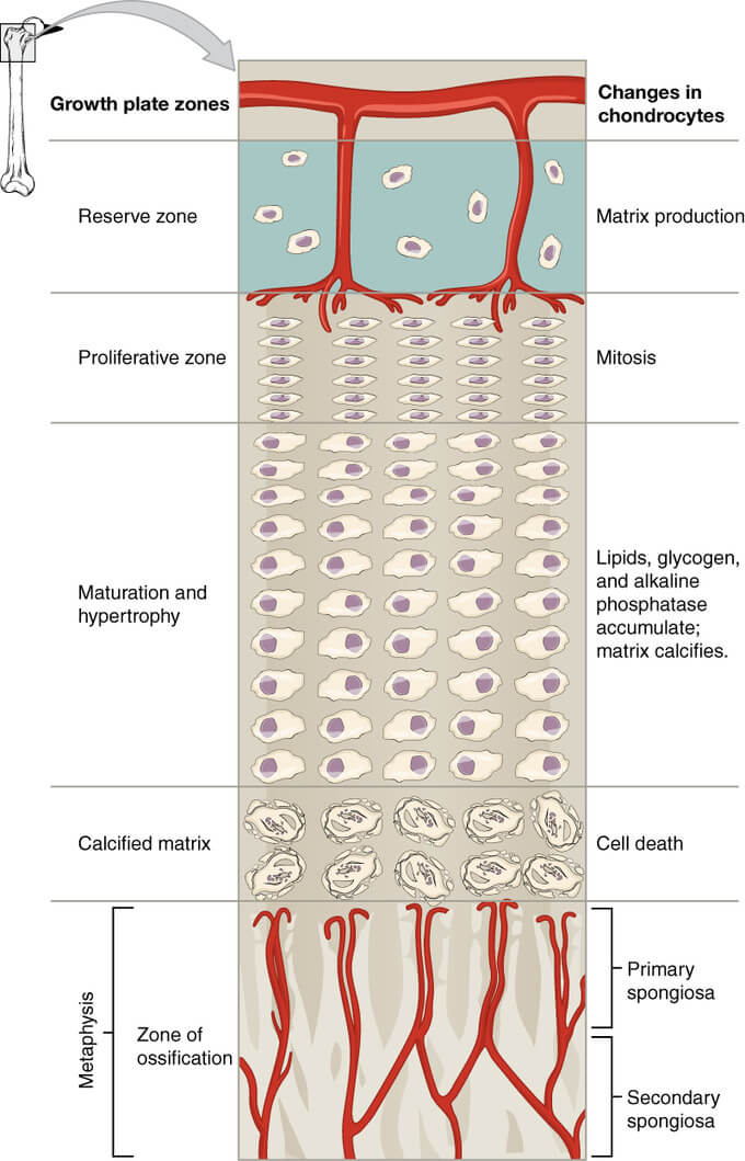
physis zones
38.8: Bone – Growth of Bone (LibreTexts libraries) [CC-BY-SA 4.0]

Zones of physis:

ZonesCharacteristicsFunctionsBlood supplyDiseases
EpiphysisEpiphyseal arteryMultiple Epiphyseal Dysplasia (MED)
Physis
1. Reserve (Resting) zoneGerminal cells of stem cell origin (responsive to hormones) surrounded by mechanically strong thick layer of matrixMatrix production

Storage
Epiphyseal arteries descend through the layer but do not supply (low oxygen tension)1. Diastrophic dysplasia
2. Pseudo-achondroplasia
3. Gaucher’s disease
2. Proliferative (Columnar) zoneStacking of chondrocytes longitudinally; surrounded by mechanically strong thick layer of matrixMatrix production

Cellular proliferation
Excellent (excellent oxygen tension)1. Achondroplasia
2. Hypo-achondroplasia
3. Gigantism
3. Hypertrophic zoneHypertrophy of chondrocytes (5-10 times) with less space for matrix (weakest layer)SCFE (non-renal failure)

Enchondroma
a. Maturation zonePreparation of matrix for calcificationLow (low oxygen tension)Mucopolysaccharidosis
b. Degenerative zonePreparation of matrix for calcificationLower (lower oxygen tension)Mucopolysaccharidosis
c. Zone of provisional calcificationProgrammed cell death of chondrocytesCalcification of matrixNil (poor oxygen tension)1. Rickets & Osteomalacia
2. Physeal fractures
MetaphysisMetaphyseal & nutrient artery branches
1. Primary spongiosaBone formationGood (good oxygen tension)1. Metaphyseal chondrodysplasia
2. Acute hematogenous osteomyelitis
3. Corner fractures
4. SCFE (renal failure)
2. Secondary spongiosaRemodelingExcellent (excellent oxygen tension)1. Osteopetrosis
2. Osteogenesis imperfecta
3. Scurvy
4. Metaphyseal dysplasia

The hypertrophic zone is therefore the weakest layer of the physis under tension, shear, and bending stress, and it is the most common area for fractures. 
  • Facebook
  • Twitter
PGMEE, MRCS, USMLE, MBBS, MD/MS AnatomyMusculoskeletal systemOrthopedics

Post navigation

Previous post
Next post

Related Posts

PGMEE, MRCS, USMLE, MBBS, MD/MS

List of Textbook for Pediatric Residents

Mar 27, 2021Mar 27, 2021

Residency in any subject in medical field is not a piece of cake. There is no limit of knowledge you need to acquire and the tougher thing is you need to acquire knowledge and skills side by side. A mastery in only one field can leave you deficient. As a…

Read More
PGMEE, MRCS, USMLE, MBBS, MD/MS gamma loop

Lower Motor Neuron Lesion (LMNL) – Anatomical Basis

Jul 25, 2016Jul 25, 2016

The anatomical basis of Upper Motor Neuron Lesion (UMNL) has already been discussed earlier. Similarly, we will explain the anatomical basis of clinical syndrome of Lower Motor Neuron Lesion (LMNL). A. Ipsilateral involvment: Lower motor neuron comprises of motor neurons in the anterior neurons and the fibers originating from them,…

Read More
PGMEE, MRCS, USMLE, MBBS, MD/MS parinaud syndrome

Parinaud Syndrome – Anatomical Basis

May 19, 2019May 19, 2019

Synonyms: Dorsal midbrain sydnrome, Pretectal syndrome Main components of Parinaud syndrome: Vertical gaze palsy Convergence-retraction nystagmus Pupillary light-near dissociation Lid retraction (Collier’s sign) Anatomical basis of Parinaud syndrome: Site of lesion: Dorsal midbrain Close to cerebral aqueduct and pineal gland – hydrocephalus and pinealoma Arterial supply – posterior cerebral artery…

Read More

Leave a Reply Cancel reply

Your email address will not be published. Required fields are marked *

This site uses Akismet to reduce spam. Learn how your comment data is processed.

Epomedicine. Anatomy of Physis (Growth Plate) [Internet]. Epomedicine; 2024 Jul 30 [cited 2026 Jul 25]. Available from: https://epomedicine.com/medical-students/anatomy-of-physis-growth-plate/.

Pre-clinical (Basic Sciences)

Anatomy

Biochemistry

Community medicine (PSM)

Embryology

Microbiology

Pathology

Pharmacology

Physiology

Clinical Sciences

Anesthesia

Dermatology

Emergency medicine

Forensic

Internal medicine

Gynecology & Obstetrics

Oncology

Ophthalmology

Orthopedics

Otorhinolaryngology (ENT)

Pediatrics

Psychiatry

Radiology

Surgery

RSS Ask Epomedicine

  • What to study for Clinical examination in Orthopedics?
  • What is the mechanism of AVNRT?

Epomedicine weekly

  • About Epomedicine
  • Contact Us
  • Author Guidelines
  • Submit Article
  • Editorial Board
  • USMLE
  • MRCS
  • Thesis
©2026 Epomedicine | WordPress Theme by SuperbThemes