Skip to content
Epomedicine

Mnemonics, Simplified Concepts & Thoughts

Epomedicine

Mnemonics, Simplified Concepts & Thoughts

hyperkalemia ecg

ECG changes in Hyperkalemia

Epomedicine, Nov 9, 2013May 26, 2019

Synonym: Hyperpotassemia

Definition: Serum potassium (K+) > 5 mEq/l

Electrophysiologic basis of ECG changes:

In patients with mild hyperkalemia, potassium conductance (IKr) through potassium channels is increased, which tend to shorten the AP duration and on the ECG causing tall tented T waves.

With severe hyperkalemia, effect on RMP becomes prominent. Increased extracellular K+ reduces the Resting Membrane Potential (RMP), i.e. makes it less negative, which leads to partial inactivation of cardiac Na+ channels resulting in slower upstroke velocity of AP. This results in impaired conduction velocity and slowing of AV conduction and intra-ventricular conduction.

a. Atria are more sensitive: Intially, PR prolongation and flattening of P waves

b. Later ventricles involved: QRS widened

c. With extreme hyperkalemia: Total inactivation of Na+ channels and heart stops (cardioplegia)

ECG features:
 
Changes in the T wave provide the earliest clues to hyperkalemia.
 
Hyperkalemia
 
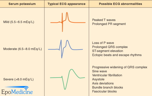
  1. At K = 5.0 to 6.0 mEq/L, rapid repolarization causes peaked T waves (best seen in leads V2 to V4).
  2. At K = 6.0 to 6.5 mEq/L, decrease in conduction causes prolonged PR and QT intervals.
  3. At K = 6.5 to 7.0 mEq/L, P waves are diminished and ST segment may be depressed.
  4. At K = 7.0 to 8.0 mEq/L, P waves disappear, QRS widens, and irregular idioventricular rhythm appears.
  5. At K = 8.0 to 10.0 mEq/L, QRS merges with T wave to produce classic sine wave (QRS-T fusion – a sinusoidal waveform).
  6. At K = 10.0 to 12.0 mEq/L, ventricular fibrillation and diastolic arrest occur.
 
Note: Hyperkalemia causes tall, peaked (tented) T waves. ECG signs may be absent if the onset of hyperkalemia is slow – as seen in chronic renal failure – even though the serum potassium is in the range of 7 – 7.5meq/L.

106 shares
  • Facebook74
  • Twitter
Emergency Medicine Cardiovascular systemECG

Post navigation

Previous post
Next post

Related Posts

Clinical Skills and Approaches

FARES method of Shoulder Reduction

Nov 30, 2019Nov 30, 2019

Indication: Anterior shoulder dislocation Position of patient: Supine Position of physician: Standing on the side of the dislocated shoulder Steps: Physician holds the wrist of the patient with both the arms keeping elbow of the patient extended and forearm in neutral position Arm is slowly abducted in brief oscillating movement…

Read More
Emergency Medicine pulmonary embolism xray

Pulmonary Embolism : Chest X-ray Signs

Oct 25, 2017

1. Classic presentation is normal X-ray in patient with dyspnea and hypoxia 2. Atelectasis or parenchymal abnormality (68%) 3. Elevated hemidiaphragm 4. Pleural effusion (Felson’s sign – pleural effusion on left > right) 5. Hampton’s hump: peripheral pleural based wedge-shaped density above the diaphragm due to pulmonary infarct 6. Westermark’s…

Read More
Emergency Medicine PE algorithm

Well’s and PERC Criteria for Pulmonary Embolism : Mnemonic

Oct 25, 2017Oct 2, 2021

Well’s Criteria (Modified and Simplified) Mnemonic: CHADS (Remember, this is not the CHADS2 score for Atrial Fibrillation) Clinical features of DVT Cancer Heart rate > 100/min Hemoptysis Alternative diagnosis less likely DVT/PE in past Surgery in past 4 weeks or Immobilization for 3 days Well’s criteria Original score Simplified score…

Read More

Leave a Reply Cancel reply

Your email address will not be published. Required fields are marked *

This site uses Akismet to reduce spam. Learn how your comment data is processed.

Pre-clinical (Basic Sciences)

Anatomy

Biochemistry

Community medicine (PSM)

Embryology

Microbiology

Pathology

Pharmacology

Physiology

Clinical Sciences

Anesthesia

Dermatology

Emergency medicine

Forensic

Internal medicine

Gynecology & Obstetrics

Oncology

Ophthalmology

Orthopedics

Otorhinolaryngology (ENT)

Pediatrics

Psychiatry

Radiology

Surgery

RSS Ask Epomedicine

  • What to study for Clinical examination in Orthopedics?
  • What is the mechanism of AVNRT?

Epomedicine weekly

  • About Epomedicine
  • Contact Us
  • Author Guidelines
  • Submit Article
  • Editorial Board
  • USMLE
  • MRCS
  • Thesis
©2026 Epomedicine | WordPress Theme by SuperbThemes