Skip to content
Epomedicine

Mnemonics, Simplified Concepts & Thoughts

Epomedicine

Mnemonics, Simplified Concepts & Thoughts

FARES method of Shoulder Reduction

Epomedicine, Nov 30, 2019Nov 30, 2019
fares method shoulder reduction

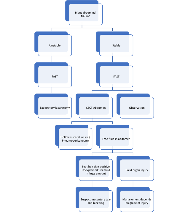
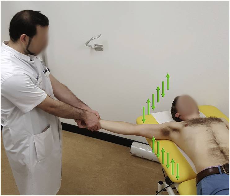
Indication: Anterior shoulder dislocation

Position of patient: Supine

Position of physician: Standing on the side of the dislocated shoulder

Steps:

  1. Physician holds the wrist of the patient with both the arms keeping elbow of the patient extended and forearm in neutral position
  2. Arm is slowly abducted in brief oscillating movement (about 5 cm up and down movement – 2 to 3 oscillations per second)
  3. After 90 degrees of abduction, add external rotation while continuing oscillations
  4. Reduction is usually achieved around 120 degrees of abduction
  5. After achieving reduction, slowly internally rotate the arm to bring the forearm to lie across the patient’s chest
https://www.youtube.com/watch?v=6DLfst2tso0

Success rate: 88-95%

Reduction time: 2-3 minutes

Pain: 1-2 out of 10 on VAS scale

Reference: Alkaduhimi H, van der Linde JA, Flipsen M, van Deurzen DF, van den Bekerom MP. A systematic and technical guide on how to reduce a shoulder dislocation. Turk J Emerg Med. 2016 Nov 18;16(4):155-168. doi: 10.1016/j.tjem.2016.09.008. PMID: 27995208; PMCID: PMC5154590.

  • Facebook
  • Twitter
Clinical Skills and Approaches Emergency Medicine Emergency medicineMusculoskeletal systemOrthopedics

Post navigation

Previous post
Next post

Related Posts

Emergency Medicine pulmonary embolism xray

Pulmonary Embolism : Chest X-ray Signs

Oct 25, 2017

1. Classic presentation is normal X-ray in patient with dyspnea and hypoxia 2. Atelectasis or parenchymal abnormality (68%) 3. Elevated hemidiaphragm 4. Pleural effusion (Felson’s sign – pleural effusion on left > right) 5. Hampton’s hump: peripheral pleural based wedge-shaped density above the diaphragm due to pulmonary infarct 6. Westermark’s…

Read More
Clinical Skills and Approaches ascitic paracentesis site

Ascitic Paracentesis : Practical Essentials

Jan 16, 2017

Absolute contraindications to Ascitic Tapping (Paracentesis) While some authors have claimed ascitic paracentesis to be free from absolute contraindications, others have listed following as absolute contraindications: Clinically evident fibrinolysis Disseminated Intravascular Coagulation (DIC) Clinically apparent oozing from needle sticks Acute abdomen requiring surgery Relative contraindications to Blind Paracentesis Ultrasound guided…

Read More
Emergency Medicine

Conus Medullaris Syndrome vs Cauda Equina Syndrome : Anatomical basis and Mnemonic

May 8, 2022May 8, 2022

Definitions Condition Vertebral level of injury Neurological level of injury ISNCI level of injury Conus Medullaris Syndrome (CMS) T12-L2 T12-S5 T11 Cauda Equina Syndrome (CES) L3-L5 L3-S5 L2 Anatomy The spinal cord ends as a tapered structure called the conus medullaris at the level of L2–L3 disc in the neonate…

Read More

Leave a Reply Cancel reply

Your email address will not be published. Required fields are marked *

This site uses Akismet to reduce spam. Learn how your comment data is processed.

Pre-clinical (Basic Sciences)

Anatomy

Biochemistry

Community medicine (PSM)

Embryology

Microbiology

Pathology

Pharmacology

Physiology

Clinical Sciences

Anesthesia

Dermatology

Emergency medicine

Forensic

Internal medicine

Gynecology & Obstetrics

Oncology

Ophthalmology

Orthopedics

Otorhinolaryngology (ENT)

Pediatrics

Psychiatry

Radiology

Surgery

RSS Ask Epomedicine

  • What to study for Clinical examination in Orthopedics?
  • What is the mechanism of AVNRT?

Epomedicine weekly

  • About Epomedicine
  • Contact Us
  • Author Guidelines
  • Submit Article
  • Editorial Board
  • USMLE
  • MRCS
  • Thesis
©2026 Epomedicine | WordPress Theme by SuperbThemes