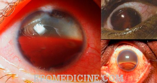
Definition: Accumulation of blood in the Anterior Chamber (AC).
Causes:
a. Post-traumatic: Trauma or surgery
b. Spontaneous:
- Neovascularization
- Ocular neoplasms
- Vascular and clotting anomalies (leukemia, hemophilia, aspirin)
Grading for Traumatic Hyphema:
- 0 : No layered blood, circulating RBCs only (Microhyphema)
- 1: Layered blood filling < 1/3 of AC
- 2: Layered blood filling 1/3 to 1/2 of AC
- 3: Layered blood filling 1/2 to less than total of AC
- 4: Total clotted blood (Blackball or 8 ball hyphema if the blood has clotted and appears black due to impaired aqueous circulation and deoxygenated blood)
Work-up:
- History and Physical examination: rule out penetrating injury, intraocular structure damage
- Intraocular pressure (IOP) measurement and dilated retinal examination
- May be: USG B scan, USG biomicroscopy, CT orbit/brain
- Sickle cell screening: African american or Mediterranean
Management:
1. Facilitate inferior settling:
- Limitation of activity with head end elevated
- Patching and shielding
2. Aropine 1% eyedrops, mild analgesics and topical steroids can be given
3. Reduce IOP:
- Antiglaucoma drugs
- AC paracentesis if uncontrolled
4. Fibrinolytic: Intracameral tPA
- For refractory or malignant raised IOP
- But risk of rebleeding
5. Surgical interventions:
Modalities:
- Hyphema evacuation with closed vitrectomy
- Paracentesis and AC washout (best to perform after 4-7 days of injury as clot has time to solidify, reducing risk of rebleed)
Indications:
- Microscopic corneal blood staining.
- Total hyphema with IOP > 30 mmHg or > 5 days
- Total hyphema or filling >75% AC for 6 days with IOP 25 mmHg or more
- >50% AC (large hyphema) for >10 days
- Sickle cell trait or Sickle cell disease with IOP > 35 mmHg for >24 hours
Sickle cell disease and hyphema:
Higher elevation in IOP (sickled RBCs cannot pass through TM)
Higher risk of central retinal artery occlusion (CRAO) and optic nerve infarction due to vascular sludging
Ocular hypotensive medications in sickle cell:
- β-blockers are safe
- Avoid CAIs (increases concentration of ascorbic acid in aqueous, decreasing pH and leading to sickling)
- Epinephrine and α-agonists (cause vasoconstriction with subsequent deoxygenation and sickling)
- Hyperosmotics (lead to hemoconcentration with vascular sludging and sickling)
Complications:
- Blood staining of cornea
- Recurrent hemorrhage
- Secondary glaucoma:
- Early: due to Trabecular meshwork obstruction, pupillary block by clot, hemolytic, steroid-induced
- Late: due to angle recession, ghost cell, PAS formation, posterior synechiae with iris bombe
- Optic atrophy
- due to traumatic optic neuropathy relating to the original injury, or glaucomatous damage secondary to increased IOP