Skip to content
Epomedicine

Mnemonics, Simplified Concepts & Thoughts

Epomedicine

Mnemonics, Simplified Concepts & Thoughts

cornea histology schematic

Histology of Cornea and Corneal Dystrophies

Epomedicine, May 22, 2014Aug 31, 2024

Prefix: kerat-

Definition: The cornea is a transparent, avascular, watch-glass like structure which forms anterior one-sixth of the outer fibrous coat of the eyeball and covers iris, pupil and anterior chamber.

Histology:

cornea histology schematic

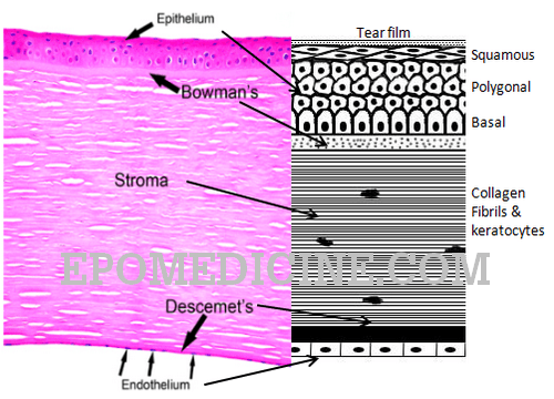
It consists of 5 distinct layers which can be remembered using the mnemonic “ABCDE“:

LayersThickness (µm)CompositionPathophysiology
Anterior epithelium50a. Top: 3-4 layers of squamous cells (uppermost are apical cells)b. Middle: 1-3 layers of wing cells (flattened polygonal shape)

c. Deep: 1 layer of basal cells

d. Basal lamina: scaffold for epithelim; collagen type IV; secreted by basal cells

Epithlial basement membrane dystrophy (AD)

Meesmann’s (AD)

Bowman’s membrane8-14Unorganized type I collagen fibers  in GAG matrix
Acellular
Reis-Buckler’s (AD)
Corneal stroma (Substantia propria)500 (~90%)~ 80% water by weightParallely organized lamellae of collagen I, IV and V in mucopolysaccharide matrix

Cells: Keratocytes, Langerhans’ cells, pigmented melanocytes, macrophages, histiocytes

Only MMP-2 is found in healthy cornea

Granular (AD)

Macular (AR)

Lattice (AD)

Descemet’s membrane10-12Anterior organized fetal banded layer (no change with age 3 µm)Posterior unorganized non-banded layer (thickens with age 2-10 µm)

PAS + true basement membrane

 
Endothelium5Single layer of interdigitating hexagonal cells (It is a misnomer as the cells are not endothelial cells)Schwalbe’s line: termination of corneal endothelium (junction between endothelium and trabecular meshwork)

Posterior embryotoxon: thickening and anterior displacement of Schwalbe’s line

Posterior polymorphous corneal dystrophy (AD)

Fuch’s dystrophy (AD)

The pre-Descemet’s layer/Dua’s layer, also termed the Dua-Fine layer and the pre-posterior limiting lamina layer, lies anterior to the Descemet’s membrane in the cornea, is 10 μm (range 6-16) thick, made predominantly of type I and some type VI collagen with abundant elastin, more than any other layer of the cornea.

Corneal Wound Healing in Short:

Epithelium: by migration and mitosis; rapid (starts 24-48 hours and completed by 6-8 days)

Stroma: non-complicated wound (avascular healing); complicated wound (vascular healing)

Descemet’s membrane and endothelium: slow regeneration

  1. Endothelium: mitosis and migration
  2. Descemet’s membrane: replacement by hyaline material derived from endothelium
12 shares
  • Facebook12
  • Twitter
PGMEE, MRCS, USMLE, MBBS, MD/MS AnatomyOphthalmology

Post navigation

Previous post
Next post

Related Posts

PGMEE, MRCS, USMLE, MBBS, MD/MS

Pharmacokinetics Mnemonics

Aug 12, 2019Aug 18, 2024

ADME is the 4-letter acronym used to describe pharmacokinetics: ABCD is another 4-letter acronym used to describe pharmacokinetics: The most important pharmacokinetic parameters from a dosing point of view are: Vd = Volume of distributionD = DoseCo = Concentration in plasmaCL = ClearancekE = Elimination constantt1/2 = Half life In…

Read More
PGMEE, MRCS, USMLE, MBBS, MD/MS hyperkalemia ecg

Hyperkalemia Management – Mnemonic Approach

Oct 24, 2020Oct 24, 2020

Mnemonic: C BIG K D Calcium gluconate (Cardiac stabilizer) It is generally accepted that calcium should be given when there are ECG changes associated with hyperkalaemia. Calcium gluconate 10% 10-30 ml IV (1-3 gm) over 5-10 minutes (Can be repeated after 5 minutes if ECG changes persistent) 0.5 ml/kg in…

Read More
PGMEE, MRCS, USMLE, MBBS, MD/MS jugular foramen

Structures passing through Jugular foramen : Mnemonic

Jun 29, 2018

Mnemonic: IPS 9,10,11 MAP JOE Anterior part: IPS Inferior Petrosal Sinus Middle part: 9, 10, 11 MAP CN IX CN X CN XI Meningeal br. of Ascending Pharyngeal artery Posterior part: JOE Jugular vein (internal; lower end of sigmoid sinus) Occipital artery Emissary vein Vernet’s syndrome (Jugular foramen syndrome) Unilateral…

Read More

Leave a Reply Cancel reply

Your email address will not be published. Required fields are marked *

This site uses Akismet to reduce spam. Learn how your comment data is processed.

Pre-clinical (Basic Sciences)

Anatomy

Biochemistry

Community medicine (PSM)

Embryology

Microbiology

Pathology

Pharmacology

Physiology

Clinical Sciences

Anesthesia

Dermatology

Emergency medicine

Forensic

Internal medicine

Gynecology & Obstetrics

Oncology

Ophthalmology

Orthopedics

Otorhinolaryngology (ENT)

Pediatrics

Psychiatry

Radiology

Surgery

RSS Ask Epomedicine

  • What to study for Clinical examination in Orthopedics?
  • What is the mechanism of AVNRT?

Epomedicine weekly

  • About Epomedicine
  • Contact Us
  • Author Guidelines
  • Submit Article
  • Editorial Board
  • USMLE
  • MRCS
  • Thesis
©2026 Epomedicine | WordPress Theme by SuperbThemes