Skip to content
Epomedicine
Epomedicine

Mnemonics, Simplified Concepts & Thoughts

  • Medical Students
  • Bedside Clinics
  • Case Reports
  • Emergency Medicine
  • Blog
  • Surgical Skills
  • Medical Mnemonics
Epomedicine

Mnemonics, Simplified Concepts & Thoughts

Shoelace technique for Delayed Primary Closure

Epomedicine, Jan 21, 2024Jan 21, 2024

Last updated on January 21, 2024

Indication: Delayed primary closure of fasciotomy wounds

Timing of Wound Closure (Primary, Secondary, Tertiary)

Sutures that can be used:

  1. Large polypropylene sutures
  2. Sialistic vessel loops
  3. Large silk sutures
  4. Ethibond
  5. Pediatric urinary catheter

Anchors: Staples (can apply 2 staples) or Metal clips

  • Interval of anchors: 1-2 cm
  • Wound to anchor distance: 0.3-0.5 cm

Knotting pattern:

The suture is attached to one side and passed through the incision to be attached on the opposite side, in a sequence that resembles a zigzag from the proximal to the distal regions – in a shoelace manner.
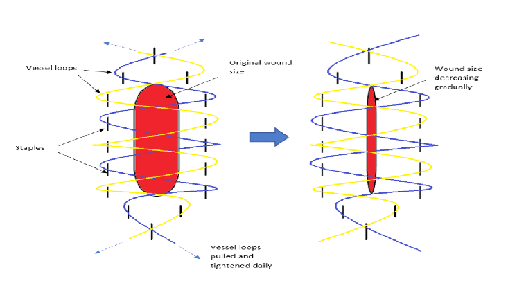
shoelace wound closure
Atan, Ahmad Arieff & Khalid, Muhammad & Chong, Fabian. (2019). Delayed reconstruction for olecranon fracture with extensive soft tissue injury and bone loss: A case report. 3. 11-16. 10.31282/joti.v2n3.47.

Tightening:

As swelling in the extremity decreases, the tension of the lacing will also decrease and redundancy in the lace will occur. During the wound inspection at the bedside, the lace is tightened using a sterile technique. After untying or cutting the knot, the lace is tightened throughout the length by stretching the suture used. Once the proper tension is reestablished, the ends of the loop are again tied snugly.

  • Rate of tightening: 24-72 hourly
  • Amount of tightening: can use capillary refill time of wound edges to guide amount of tightening

Time to delayed primary closure: 1-4 weeks (depending on wound)

  • After several days, the wound edges approximate, and it is usually possible to perform delayed primary closure with non-absorbable suture.

Complications:

  1. Infection
  2. Postoperative retractile scarring
  3. Partial skin necrosis

References:

  1. Obuh OO, Esomu EO, Sydney RO. Suturing Dermatotraction Techniques in Closing Fasciotomy Wounds: A Systematic Review. Cureus. 2023 Apr 13;15(4):e37550. doi: 10.7759/cureus.37550. PMID: 37197103; PMCID: PMC10184723.
  2. Berman, S. S., Schilling, J. D., McIntyre, K. E., Hunter, G. C., & Bernhard, V. M. (1994). Shoelace technique for delayed primary closure of fasciotomies. The American Journal of Surgery, 167(4), 435–436. doi:10.1016/0002-9610(94)90130-9 
2 shares
  • Facebook
  • Twitter
Surgical Skills OrthopedicsSurgical skills

Post navigation

Previous post
Next post

Leave a Reply Cancel reply

Your email address will not be published. Required fields are marked *

This site uses Akismet to reduce spam. Learn how your comment data is processed.

Epomedicine. Shoelace technique for Delayed Primary Closure [Internet]. Epomedicine; 2024 Jan 21 [cited 2025 May 9]. Available from: https://epomedicine.com/surgical-skills/shoelace-technique-for-delayed-primary-closure/.

Pre-clinical (Basic Sciences)

Anatomy

Biochemistry

Community medicine (PSM)

Embryology

Microbiology

Pathology

Pharmacology

Physiology

Clinical Sciences

Anesthesia

Dermatology

Emergency medicine

Forensic

Internal medicine

Gynecology & Obstetrics

Oncology

Ophthalmology

Orthopedics

Otorhinolaryngology (ENT)

Pediatrics

Psychiatry

Radiology

Surgery

RSS Ask Epomedicine

  • What to study for Clinical examination in Orthopedics?
  • What is the mechanism of AVNRT?

Epomedicine weekly

  • About Epomedicine
  • Contact Us
  • Author Guidelines
  • Submit Article
  • Editorial Board
  • USMLE
  • MRCS
  • Thesis
©2025 Epomedicine . All rights reserved.