Skip to content
Epomedicine

Mnemonics, Simplified Concepts & Thoughts

Epomedicine

Mnemonics, Simplified Concepts & Thoughts

Radiological Lines for assessing Basilar Invagination : Mnemonic

Epomedicine, Nov 24, 2024Feb 21, 2025

Mnemonic: Imagine a letter “Z“.
a. McRae’s line has “R” and is at Roof.
b. Chamberlain’s line starts with “C” and is at Center.
c. McGregor’s line has “G” and is at Ground.

basal skull lines

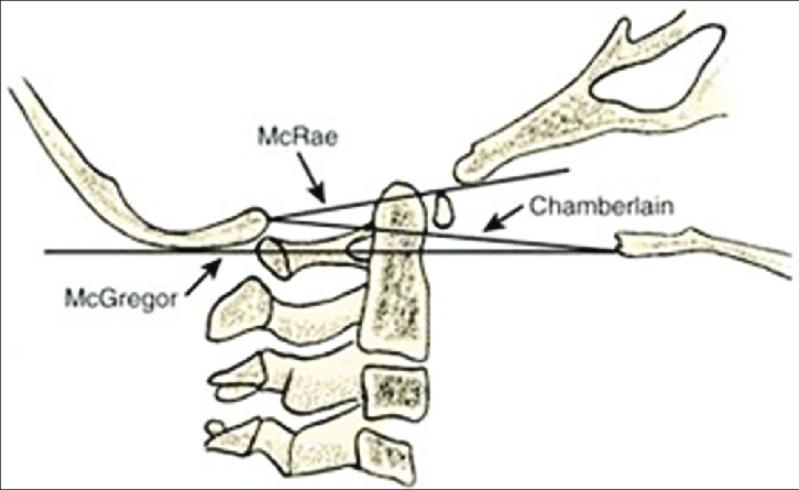
a. McRae’s line (foramen magnum line): drawn from anterior margin of foramen (basion) to posterior margin of foramen (opisthion)

  • Normal: Tip of dens should not cross this line

b. Chamberlain’s line: drawn from posterior end of hard palate to posterior margin of foramen (opisthion)

  • Normal: Tip of dens is no more than 3 mm above this line

c. McGregor’s line: drawn from posterior end of hard palate to posterior occiput curve

  • Normal: Tip of dens is no more than 4.5 mm above this line

Other lines

ranawat and redlund line

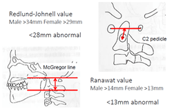
a. Wackenheim’s line (Clivus canal line): Line drawn along clivus into cervical canal

  • Normal: Tip of dens is ventral and tangential to this line

b. Ranawat’s line: A perpendicular line drawn from center of C2 pedicle towards a line from center of anterior to posterior arch of C1

  • Abnormal: <13 mm

c. Redlund-Johnell’s method: Measure distance between midpoint of C2 body inferior margin and McGregor’s line

  • Abnormal: <28 mm
  • Facebook
  • Twitter
PGMEE, MRCS, USMLE, MBBS, MD/MS Nervous systemOrthopedicsRadiology

Post navigation

Previous post
Next post

Related Posts

PGMEE, MRCS, USMLE, MBBS, MD/MS

Pes Anserinus : Mnemonic

Apr 29, 2021Apr 29, 2021

Pes Anserinus is composed of the combination of tendinous insertions of the sartorius, gracilis and semitendinosus muscles (guy ropes muscles) which attaches to the medial side of tibia to generate a “goose’s foot” like appearance. These three muscles are mainly flexors of the knee but also have a role in…

Read More
PGMEE, MRCS, USMLE, MBBS, MD/MS

Ligaments of Pelvis

Oct 3, 2020Oct 3, 2020

Inherent stability of the pelvis is provided by ligaments. The 3 groups of ligaments are: 1. Sacrum to Pelvis: Sacroiliac ligamentous complex: is divided into posterior (short and long) and anterior ligaments. Posterior ligaments provide most of the stability. Sacrotuberous ligament: runs from the posterolateral aspect of the sacrum and the…

Read More
PGMEE, MRCS, USMLE, MBBS, MD/MS

Paget’s Disease of bone : Mnemonic

Jul 20, 2022Jul 20, 2022

Synonyms: Osteitis deformans Collage of matrix madness Mnemonic: Sixteen “P”s 1. Paramyxovirus infection and inclusions in osteocalsts 2. Pain (most common symptom – localized fractures or nerve compressions) 3. Pelvis (most common site) 4. Polyostotic (more common; 85%) 5. Phases: Active (lytic, mixed, sclerotic) and Inactive 6. Plasma volume expansion…

Read More

Leave a Reply Cancel reply

Your email address will not be published. Required fields are marked *

This site uses Akismet to reduce spam. Learn how your comment data is processed.

Pre-clinical (Basic Sciences)

Anatomy

Biochemistry

Community medicine (PSM)

Embryology

Microbiology

Pathology

Pharmacology

Physiology

Clinical Sciences

Anesthesia

Dermatology

Emergency medicine

Forensic

Internal medicine

Gynecology & Obstetrics

Oncology

Ophthalmology

Orthopedics

Otorhinolaryngology (ENT)

Pediatrics

Psychiatry

Radiology

Surgery

RSS Ask Epomedicine

  • What to study for Clinical examination in Orthopedics?
  • What is the mechanism of AVNRT?

Epomedicine weekly

  • About Epomedicine
  • Contact Us
  • Author Guidelines
  • Submit Article
  • Editorial Board
  • USMLE
  • MRCS
  • Thesis
©2026 Epomedicine | WordPress Theme by SuperbThemes