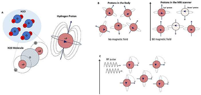
Human body is made up of water, which means a large number of atoms inside our body is hydrogen atoms, the nucleus of which contains a positively charged proton that spins (or precesses) around an axis like a child’s top. This spinning generates its own tiny magnetic field, giving the proton its own north and south poles. Under normal circumastances, these protons spin on randomly oriented axis.
When magnetic field is turned on, the axes align with the more powerful external magnetic field with some aligning parallel (low energy state) to the field and some aligning anti-parallel (high energy state) to magnetic field, cancelling each other. The larger the external magnetic field, the greater the difference in energy levels and the larger the excess number aligned with the field.
A radiofrequency (RF) coil is used to send pulses is applied for a few milliseconds, excess protons aligning parallel to the magnetic field flips their configuration to high energy state (antiparallel). When the RF pulse stops:
- Absorbed RF energy is released by protons
- T1 relaxation (recovery): recovery of longitudinal orientation; T1 time refers to interval where 63% of longitudinal magnetization is recovered
- T2 relaxation (dephasing): loss of transverse magnetization; T2 time refers to time where only 37% of original transverse magnetization is present
In doing so, a signal is emitted which gets turned into an electric current, which the scanner digitizes. The lower the water content in an area, the fewer hydrogen protons there will be emitting signals back to the RF coils. In absolute terms, in biologic tissues, T1 is about 300 to 2000 msec, and T2 is about 30 to 150 msec (T1 is longer than T2; T1 is always longer than T2 except in pure water in which T1=T2).
In T1-weighted images, fat, e.g. bone marrow, has a bright signal; on T2 weighted images, fluid has a bright signal. Tissues with little fat or water, e.g. cortical bone, ligaments and tendons, are dark on both T1- and T2 weighted images. T1 is good for anatomy (anatomy scans) as it is weighted towards fat, while T2 is good for pathology (pathology scans) as it is weighted towards water. Pathologic tissues tend to have higher water content.
Mnemonic:
a. Long drink: Fluids have long T1 and long T2
b. Fast food (hamburgers): Fats have shorter T1 and shorter T2 compared to fluids
c. In T1, only 1 thing is bright (fat is bright)
d. In T2, 2 things are bright (fat and water both are bright)
| Tissue | Signal on T1 weighted image | Signal on T2 weighted image |
| Fluid (CSF, Urine) | High | High |
| Fat | High | Low |
| Muscle | Intermediate | Intermediate |
| Cartilage | Low | High |
| Cortical bone | Low | Low |
Grey mater has a higher water content, i.e. contains more protons, its signal intensity is higher than that of the white mater.
Paramagnetic substances, e.g. the contrast medium Gadolinium-DTPA, or even deoxyglobin and methemoglobin found in hematomas shorten T1 and T2 of the surrounding protons. This results in a signal increase in T1-weighted images and a signal decrease in T2-weighted images. Enhancement following contrast injection is due to accumulation of contrast (mostly due to leaky blood vessels) in cases of tumors, areas of inflammation, or infection.
T1-weighted images are the predominant imaging technique used after contrast medium injection.
Fat suppression (or attenuation or saturation) is performed in T1 weighted sequences, to suppress the bright signal from fat. This is performed after administration of gadolinium contrast to better appreciate enhancing tissue or to prove if the particular tissue is fatty. In T2 images, fat suppression using STIR (or other methods) can help in appreciation of edema in fatty tissues. Similarly, fluid suppression using FLAIR can help in appreciation of parenchymal edema by supressing high signal intensity of CSF.
T1 images and T2 FLAIR can be differentiated. T1 sequences will have grey matter being darker than white matter. T2 weighted sequences, whether fluid attenuated or not, will have white matter being darker than grey matter.