Skip to content
Epomedicine

Mnemonics, Simplified Concepts & Thoughts

Epomedicine

Mnemonics, Simplified Concepts & Thoughts

Linea Aspera

Epomedicine, Oct 26, 2022May 14, 2024

Linea aspera is a ridge of roughened surface on the posterior surface of the shaft of femur which serves as a site for attachment of muscles and intermuscular septum.

femur
“Femur, anterior and posterior views with labels – Appendicular Skeleton Visual Atlas, page 22” by Rob Swatski is licensed under CC BY-NC 2.0.

The various structures attached to linea aspera can be remembered using few mnemonics:

Mnemonic 1#: 1, 2, 3

It gives attachment to:

  1. One biceps femoris (short head)
  2. Two vasti (vastus medialis and vastus lateralis)
  3. Three adductors (adductor longus, adductor brevis and adductor magnus) and Three intermuscular septum
Linea aspera schematic
Schematic diagram of Linea aspera; M: Medial; L: Lateral

Mnemonic 2#:

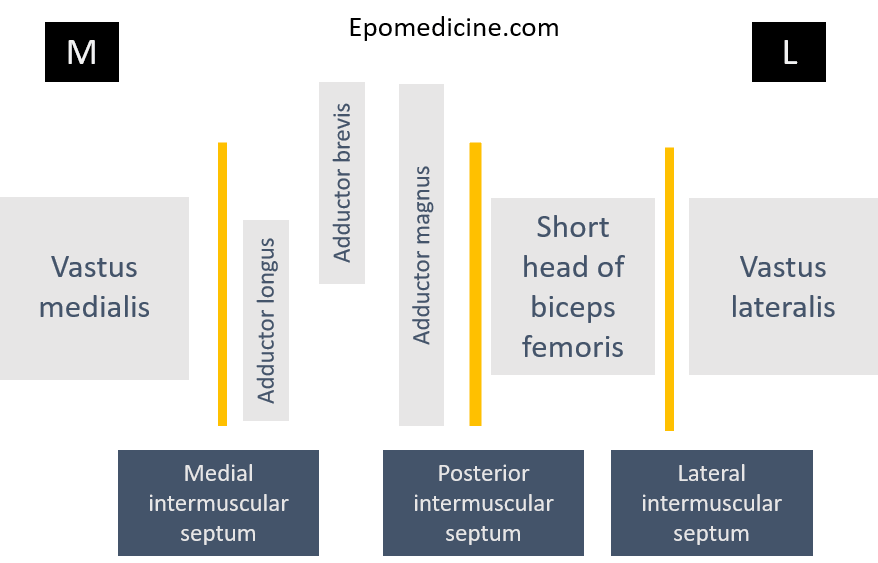
1. The area medial and lateral of medial and lateral intermuscular septum respectively is the anterior compartment.

  • Medial most attachment: Vastus medialis
  • Lateral most attachment: Vastus lateralis

2. The area between medial intermuscular septum and posterior intermuscular septum is the medial compartment. The arrangement of muscles from medial to lateral can be remembered using the mnemonic: ALABAMa

  • Adductor Longus (inferior)
  • Adductor Brevis (superior)
  • Adductor Magnus

3. The area between posterior intermuscular septum and lateral intermuscular septum is the posterior compartment. Only 1 muscle of posterior compartment of thigh arises from femur, i.e.:

  • Biceps femoris (short head)
  • Facebook
  • Twitter
PGMEE, MRCS, USMLE, MBBS, MD/MS AnatomyMusculoskeletal system

Post navigation

Previous post
Next post

Related Posts

PGMEE, MRCS, USMLE, MBBS, MD/MS

Renal Tubular Acidosis (RTA) : Mnemonics

Nov 18, 2020Nov 18, 2020

Renal Tubular Acidosis (RTA) cause non-anion gap metabolic acidosis. Type 1: H+ excretion defect (A proton or 1st element of periodic table) This occurs in distal tubule (hence, distal defect) K+ is excreted instead of H+ causing Hypokalemia. Distal tubule H+ is non-functioning – urine pH >5.5. Chronic acidosis leads…

Read More
PGMEE, MRCS, USMLE, MBBS, MD/MS

Meckel’s Diverticulum : Rule of 2

Sep 7, 2023Sep 7, 2023

General description Rule of 2s 1. Occurs in 2% population 2. Symptomatic cases are 2 times more common in males 3. Located within 2 feet proximal to ileocecal valve 4. 2 inches long and 2 cm diameter 5. 2 types of ectopic mucosa: gastric and pancreatic 6. Symptomatic only in…

Read More
PGMEE, MRCS, USMLE, MBBS, MD/MS air bronchogram

Chest X-ray: Alveolar vs Interstitial Disease

Jun 19, 2015Jun 19, 2015

Interstitium is the scaffolding that supports the alveolar walls and surrounds both the alveoli and the terminal bronchioles. Neither alveoli nor interstitium is visible on a chest X-ray when normal. It is necessary to analyze whether the pattern of diffuse opacification in the lung field is alveolar or interstitial. Terms:…

Read More

Leave a Reply Cancel reply

Your email address will not be published. Required fields are marked *

This site uses Akismet to reduce spam. Learn how your comment data is processed.

Pre-clinical (Basic Sciences)

Anatomy

Biochemistry

Community medicine (PSM)

Embryology

Microbiology

Pathology

Pharmacology

Physiology

Clinical Sciences

Anesthesia

Dermatology

Emergency medicine

Forensic

Internal medicine

Gynecology & Obstetrics

Oncology

Ophthalmology

Orthopedics

Otorhinolaryngology (ENT)

Pediatrics

Psychiatry

Radiology

Surgery

RSS Ask Epomedicine

  • What to study for Clinical examination in Orthopedics?
  • What is the mechanism of AVNRT?

Epomedicine weekly

  • About Epomedicine
  • Contact Us
  • Author Guidelines
  • Submit Article
  • Editorial Board
  • USMLE
  • MRCS
  • Thesis
©2026 Epomedicine | WordPress Theme by SuperbThemes