Skip to content
Epomedicine

Mnemonics, Simplified Concepts & Thoughts

Epomedicine

Mnemonics, Simplified Concepts & Thoughts

hypertensive retinopathy

Hypertensive Retinopathy Grading – Simplified

Epomedicine, Jun 1, 2019Jun 1, 2019

Hypertensive retinopathy has been classified by Keith, Wagener & Barker and Scheie (hypertensive and arteriosclerotic features). With this mnemonic, we will have a grading with combination of all these classifications.
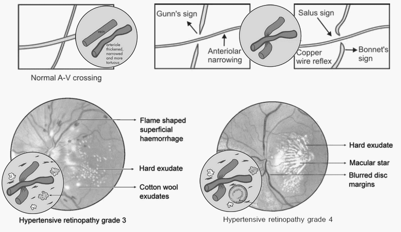
hypertensive retinopathy
Hypertensive retinopathy changes – schematic

Mnemonic: SAFEs

I – Slight Systemic (generalized) narrowing of arterioles

II – AV deflection (at Angle) and focal narrowing of arteriorles

  • Angled deflection of vein at AV crossing: Salu’s sign

III – Flame hemorrhages, Flame color arterioles and Frank AV crossing changes

  • Frank AV crossing changes:
    • Bonnet sign: Banking of veins at AV crossing
    • Marcus-gunn sign: Tapering of veins at AV crossing
  • Flame color arterioles: Copper wiring
  • Flame shaped hemorrhages, Cotton wool exudates and hard exudates

Mnemonic for Eponymous AV crossing changes:

Salu’s sign: “S” like venous deflection at AV crossing
Bonnet sign: “B” for Banking of vein distal to AV crossing
Gunn sign:

IV – Edema and Exudates surrounding macula + silver wiring

  • Edema: Papilledema
  • Exudates surrounding macula: Macular star
20 shares
  • Facebook20
  • Twitter
PGMEE, MRCS, USMLE, MBBS, MD/MS Emergency medicineInternal medicineOphthalmology

Post navigation

Previous post
Next post

Related Posts

Applied anatomy of Knee Joint

Jan 5, 2014

A. Osteology: 1. Femur: Largest bone in the body Distal femur possess 2 condyles of which the medial one is larger Medial epicondyle is more porminent and supports the adductor tubercle 2. Patella (Knee cap): Largest sesamoid bone Functions: fulcrum for quadriceps protects knee joint enhances lubrication and nutrition of…

Read More
PGMEE, MRCS, USMLE, MBBS, MD/MS

Surface Anatomy of Pleura and Lung

May 19, 2024

Surface Anatomy of Pleura Mnemonic: 2, 4, 6, 8, 10, 12 rule 1. Starts about 2 cm above the midpoint of medial 1/3 of clavicle. 2. Meet in the midline at rib 2. 3. Left side reaches sternal line at rib 4 (to make room for heart). 4. Right side…

Read More
PGMEE, MRCS, USMLE, MBBS, MD/MS hip deformity

Hip and Shoulder Deformities

Jun 7, 2019Jun 7, 2019

HIP Flexion, Abduction, External rotation (FABER) + apparent lengthening: Synovitis Flexion, Abduction, External rotation (FABER) + true lengthening: Anterior dislocation of hip (Obturator) Extension+ Abduction + External rotation (EABER): Anterior dislocation of hip (Superior or Pubic) Flexion, Adduction, Internal rotation (FADIR) + true shortening: Arthritis, Posterior dislocation of hip Flexion,…

Read More

Leave a Reply Cancel reply

Your email address will not be published. Required fields are marked *

This site uses Akismet to reduce spam. Learn how your comment data is processed.

Pre-clinical (Basic Sciences)

Anatomy

Biochemistry

Community medicine (PSM)

Embryology

Microbiology

Pathology

Pharmacology

Physiology

Clinical Sciences

Anesthesia

Dermatology

Emergency medicine

Forensic

Internal medicine

Gynecology & Obstetrics

Oncology

Ophthalmology

Orthopedics

Otorhinolaryngology (ENT)

Pediatrics

Psychiatry

Radiology

Surgery

RSS Ask Epomedicine

  • What to study for Clinical examination in Orthopedics?
  • What is the mechanism of AVNRT?

Epomedicine weekly

  • About Epomedicine
  • Contact Us
  • Author Guidelines
  • Submit Article
  • Editorial Board
  • USMLE
  • MRCS
  • Thesis
©2026 Epomedicine | WordPress Theme by SuperbThemes