Skip to content
Epomedicine

Mnemonics, Simplified Concepts & Thoughts

Epomedicine

Mnemonics, Simplified Concepts & Thoughts

Elbow ligaments : Simplified Anatomy

Epomedicine, Apr 7, 2020Apr 7, 2020
Elbow ligaments

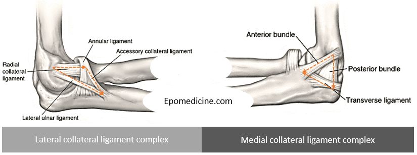
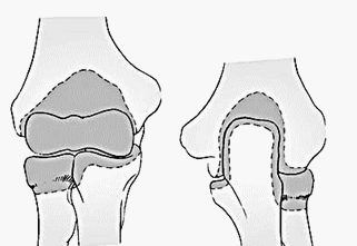
Lateral collateral ligament complex

Restraint to varus and posterolateral rotatory instability.

Anatomy is more variable.

LCL arises from lateral humeral condyle at a point through which the axis of rotation passes – it maintains a uniform tension throughout the arc of motion.

Annular ligament is a “U” shaped ligament that attaches to anterior and posterior portion of sigmoid notch (radial notch) of proximal ulna and encircles the radial head.

  • Anterior insertion: taut in supination
  • Posterior insertion: taut in pronation

The 3 other ligaments form a triangle with 3 points (lateral epicondyle, supinator crest of proximal-lateral ulna, annular ligament):

  1. Radial collateral ligament (RCL): Lateral epicondyle to Annular ligament
    • Dimension: 20 X 8 mm
  2. Lateral ulnar collateral ligament (LUCL): Lateral epicondyle to Supinator crest
  3. Accesory lateral collateral ligament: Annular ligament to Supinator crest
    • Present in 33% population
    • Stabilizes annular ligament

Key varus stabilizer: Lateral ulnar collateral ligament (LUCL)

Medial collateral ligament complex

Restraint to valgus and posteromedial rotatory instability.

Anterior bundle attaches inferior to axis of rotation (always taut) and Posterior bundle attaches posterior and inferior to axis of rotation (taut in flexion >90 degrees).

The 3 ligaments form a triangle with 3 points (medial epicondyle, medial coronoid process and medial olecranon):

  1. Anterior bundle (anterior oblique): Medial epicondyle to medial coronoid process (sublime tubercle)
    • 4 ot 5 mm width
  2. Posterior bundle (bardinet ligament): Medial epicondyle to olecranon (sigmoid notch)
    • 5 to 6 mm width
  3. Transverse bundle (ligament of cooper): Medial olecranon to medial coronoid process

Key valgus stabilizer: Anterior bundle of MCL

Other ligaments

  1. Quadrate ligament: Anterolateral ulna to Anterior radial neck (under annular ligament).
    • Tight in supination, stabilizes joint during prono-supination
  2. Oblique cord: Proximal lateral ulna Radial neck
    • Prevents downward movement of radius, stabilizes joint during prono-supination

Capsule

elbow capsule

Anterior capsule:

  • Proximally: above coronoid and radial fossa
  • Distally: coronoid medially and annular ligament laterally

Posterior capsule:

  • Proximally: just above olecranon fossa
  • Distally: medial and lateral articular margin of sigmoid notch

Greatest capacity of elbow occurs at 80 degrees flexion and is 25-30 ml. Capsule is a secondary stabilizer.

19 shares
  • Facebook19
  • Twitter
PGMEE, MRCS, USMLE, MBBS, MD/MS AnatomyMusculoskeletal systemOrthopedics

Post navigation

Previous post
Next post

Related Posts

PGMEE, MRCS, USMLE, MBBS, MD/MS eye schematic

Embryology of Eye : Mnemonic

Mar 3, 2019Mar 3, 2019

Surface ectoderm derivatives Mnemonic: LEVeL Lens Epithelium All ocular adnexa including mebomian gland and glands of Zeis and Moll Skin Cornea Conjunctiva Vitreous (portion) Lacrimal glands and drainage system Neuro-ectoderm (Optic vesicle and cup) derivatives Mnemonic: MORE Muscles of pupil (constrictor and dilator pupillae) Optic nerve Retinal pigment epithelium Epithelium…

Read More
PGMEE, MRCS, USMLE, MBBS, MD/MS

Heart Sounds – Abnormal

Apr 24, 2019May 23, 2020

The basic clinical examination of the Cardiac System is by Auscultation (listening to the sounds made by the heart). As explained in the previous article physiological sounds made in each cardiac cycle viz. S1 and S2 due to the closure of the Atrio-Ventricular Valves and Outlet Valve in synchrony. Please…

Read More
PGMEE, MRCS, USMLE, MBBS, MD/MS medulla drawing

How to draw Medulla Oblongata Cross-section ?

Jul 24, 2016Jul 24, 2016

Like in Midbrain and Pons: Corticospinal tract passes ventrally Ventricular system is located dorsally in midline Cranial nerve nuclei are located just anterior to the ventricle Medial longitudinal fasciculus is present around the center Another important thing to remember is that, the caudal medulla resembles “spinal cord“: Circular in shape…

Read More

Leave a Reply Cancel reply

Your email address will not be published. Required fields are marked *

This site uses Akismet to reduce spam. Learn how your comment data is processed.

Pre-clinical (Basic Sciences)

Anatomy

Biochemistry

Community medicine (PSM)

Embryology

Microbiology

Pathology

Pharmacology

Physiology

Clinical Sciences

Anesthesia

Dermatology

Emergency medicine

Forensic

Internal medicine

Gynecology & Obstetrics

Oncology

Ophthalmology

Orthopedics

Otorhinolaryngology (ENT)

Pediatrics

Psychiatry

Radiology

Surgery

RSS Ask Epomedicine

  • What to study for Clinical examination in Orthopedics?
  • What is the mechanism of AVNRT?

Epomedicine weekly

  • About Epomedicine
  • Contact Us
  • Author Guidelines
  • Submit Article
  • Editorial Board
  • USMLE
  • MRCS
  • Thesis
©2026 Epomedicine | WordPress Theme by SuperbThemes