Several radiological classifications have been developed to describe the long-term changes in the hip following septic arthritis and to help guide management.
- Hunka proposed one of the earliest systems, dividing cases into five types based on findings from just 10 patients followed for 11 years.
- Building on this, Choi and colleagues refined Hunka’s classification using data from 34 affected hips in 31 children.
- More recently, Forlin and Milani reviewed 41 hips in 37 patients, categorizing them primarily by the relationship between the femur and acetabulum, with further subgroups according to proximal femoral morphology.
- Johari’s classification focuses on 2 key practical features: joint stability and whether the capital femoral epiphysis is present or absent.
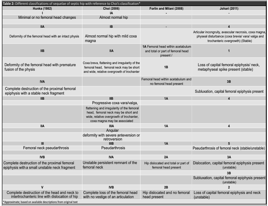
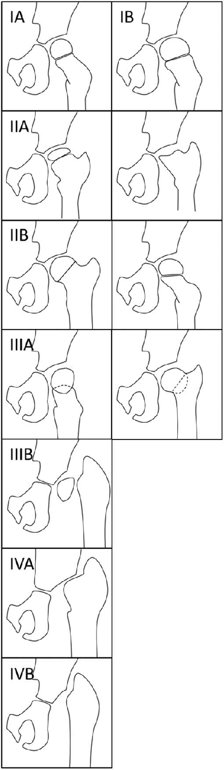
Hunka classification
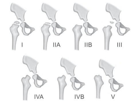
- Type I: Absent or minimal femoral head changes
- Type II
- IIa: Deformity of the femoral head with an intact physis
- IIb: Deformity of the femoral head with premature physeal closure
- Type III: Pseudoarthrosis of the femoral neck
- Type IV
- IVa: Complete destruction of the capital femoral epiphysis with a stable neck fragment
- IVb: Complete destruction of the capital femoral epiphysis with a small unstable neck fragment
- Type V: Complete destruction of both the femoral head and the femoral neck to the intertrochanteric line with hip dislocation
Choi classification
- Type I:
- Ia- Normal radiograph
- Ib- Avascular necrosis (Early Perthes like)
- Type II: Involvement of the epiphysis, physis, and metaphysis – results from avascular necrosis, with or without significant damage to the capital femoral physis.
- IIa- Coxa breva (Symmetric closure of physis)
- IIb- Coxa vara/Coxa valga (Asymmetric closure of physis)
- Type III: Damage to the femoral neck
- IIIa- Coxa vara, coxa valga, with or without excessive femoral ante version or retroversion
- IIIb- Pseudarthrosis of the femoral neck
- Type IV: Loss of the femoral head/neck
- IVa – Segment of the femoral neck is preserved
- IVb – No femoral neck remnant
Forlin and Milani classification
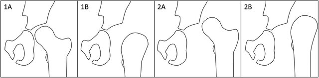
Grade 1: Hips with the head or the femoral neck within the acetabulum
- A: Femoral head present (complete or partial)
- B: Femoral head absent
Grade 2: Hips are dislocated
- A: Femoral head present
- B: Femoral head absent
Johari classification
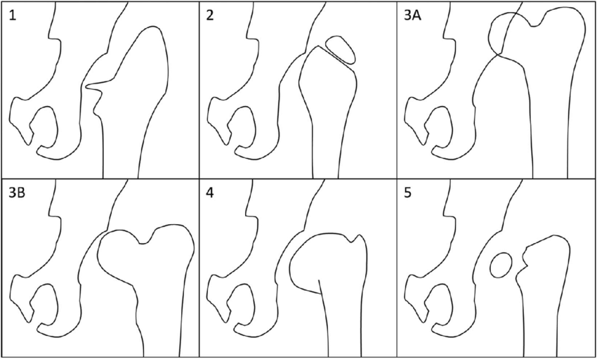
- Group 1: Loss of capital femoral epiphysis (CFE)/neck, metaphyseal spike present, stable.
- Group 2: Loss of CFE and neck, unstable.
- Group 3:
- Group 3A: Dislocation, CFE present, unstable.
- Group 3B: Subluxation, CFE present, unstable.
- Group 4: Articular incongruity, avascular necrosis, coxa magna, physeal disturbance (coxa breva, coxa vara, coxa valga, and trochanteric overgrowth), stable.
- Group 5: Pseudarthrosis of the femoral neck, stable/unstable.
References:
- Kumar Singh A, Gupta P, Kamath S, Moturu D, Reddy J, Moka S, Jethwa R, Shail S, Ganjwala D, Shah H. Reliability of Radiologic Classifications of Sequelae of Septic Arthritis of the Hip in Children. J Pediatr Orthop. 2024 Oct 1;44(9):e838-e845. doi: 10.1097/BPO.0000000000002758. Epub 2024 Jun 20. PMID: 38898555.
- Sequelae Of Septic Arthritis Of The Hip
- 10- Article_symposium 3_IJPO 2020.cdr