Skip to content
Epomedicine

Mnemonics, Simplified Concepts & Thoughts

Epomedicine

Mnemonics, Simplified Concepts & Thoughts

BIND score in severe hyperbilirubinemia

Dr. Pedchrome, Oct 27, 2022Oct 27, 2022

Bilirubin-induced neurologic dysfunction (BIND) Score is used to assess bilirubin induced encephalopathy in neonates with severe hyperbilirubinemia. Johnson et al developed the BIND score to help identify an infant who requires more aggressive monitoring and management.

Kernicterus MRI
Magnetic resonance imaging of the head. Hyperintense basal ganglia lesions on T2-weighted images [Gladys Cossio de Gurrola et al., CC BY 2.0, via Wikimedia Commons]

The scoring system has 3 parameters:

1. Cry pattern

2. Behavior and mental status

3. Muscle tone

bind scoring

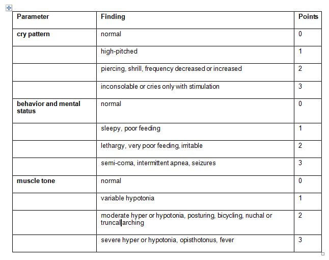
BIND score = Total score (Points for all 3 parameters scored separately and added)

Interpretation:

  • minimum score: 0
  • maximum score: 9
BIND ScoreStage
1 to 3Stage 1A
4 to 6Stage 1B
7 to 9Stage II

Stages of BIND:

Stage of BINDFeatures
IAminimal signs; totally reversible with therapy
IBprogressive signs but reversible with therapy
IIirreversible signs but severity decreased with prompt and aggressive therapy

References:

Johnson L, Brown AK, Bhutani VK. BIND – A clinical score for bilirubin induced neurologic dysfunction in newborns. Pediatrics. 1999; 104 (Supplement): 746-747.

http://www.meducator3.net/algorithms/content/clinical-severity-acute-bilirubin-induced-neurologic-dysfunction-bind-score

Resource for reading: http://pediatrics.aappublications.org/content/134/5/e1330 

Pediatric doctor
Dr. Pedchrome

MD Pediatrics and Fellowship Neonatology, he chooses to stay anonymous. He often writes his views online as well as share few important topics for medical students, doctors and specially parents. He does research in pediatrics.

  • Facebook
  • Twitter
PGMEE, MRCS, USMLE, MBBS, MD/MS NeonatesPediatrics

Post navigation

Previous post
Next post

Related Posts

PGMEE, MRCS, USMLE, MBBS, MD/MS

Crowe Classification in Adult Hip Dysplasia

Jun 4, 2025Jun 4, 2025

Crowe Classification Class I: <50% subluxation or proximal displacement <10% of pelvic height Class II: 50-75% subluxation or proximal displacement 10-15% of pelvic height Class III: 75-100% subluxation or proximal displacement 15-20% of pelvic height Class IV: >100% subluxation or proximal displacement >10% of pelvic height and deficient true acetabulum…

Read More
PGMEE, MRCS, USMLE, MBBS, MD/MS parinaud syndrome

Parinaud Syndrome – Anatomical Basis

May 19, 2019May 19, 2019

Synonyms: Dorsal midbrain sydnrome, Pretectal syndrome Main components of Parinaud syndrome: Vertical gaze palsy Convergence-retraction nystagmus Pupillary light-near dissociation Lid retraction (Collier’s sign) Anatomical basis of Parinaud syndrome: Site of lesion: Dorsal midbrain Close to cerebral aqueduct and pineal gland – hydrocephalus and pinealoma Arterial supply – posterior cerebral artery…

Read More
PGMEE, MRCS, USMLE, MBBS, MD/MS lateral brainstem syndrome

Anatomical basis of Wallenberg (Lateral Medullary) Syndrome : Mnemonic

Jan 21, 2016Oct 26, 2022

Before proceeding into the disease itself, let’s review – relevant anatomy of the medulla with a simple mnemonic. The Side (lateral) part of Medulla contains 6 “S“ 1. Spinocerebellar tract Posterior spinocerebellar tract: Ascends and enters to ipsilateral cerebellum via ipsilateral inferior cerebellar peduncle Anterior spinocerebellar tract: Ascends and enters…

Read More

Leave a Reply Cancel reply

Your email address will not be published. Required fields are marked *

This site uses Akismet to reduce spam. Learn how your comment data is processed.

Pre-clinical (Basic Sciences)

Anatomy

Biochemistry

Community medicine (PSM)

Embryology

Microbiology

Pathology

Pharmacology

Physiology

Clinical Sciences

Anesthesia

Dermatology

Emergency medicine

Forensic

Internal medicine

Gynecology & Obstetrics

Oncology

Ophthalmology

Orthopedics

Otorhinolaryngology (ENT)

Pediatrics

Psychiatry

Radiology

Surgery

RSS Ask Epomedicine

  • What to study for Clinical examination in Orthopedics?
  • What is the mechanism of AVNRT?

Epomedicine weekly

  • About Epomedicine
  • Contact Us
  • Author Guidelines
  • Submit Article
  • Editorial Board
  • USMLE
  • MRCS
  • Thesis
©2026 Epomedicine | WordPress Theme by SuperbThemes