Propensity
- Subarticular regions:
- Lie at the most distant parts of the body’s vascular territory
- Largely enclosed by cartilage, giving restricted access to blood vessels
- Endarterioles with limited collateral connections
- Marrow and bone cells:
- Vascular sinusoids unlike arterial capillaries have no adventitial layer and their patency is determined by the volume and pressure of the surrounding marrow tissue
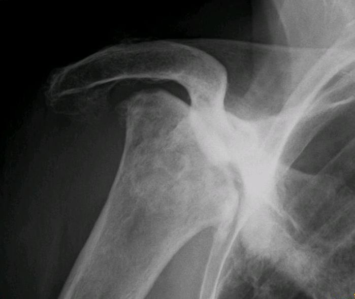
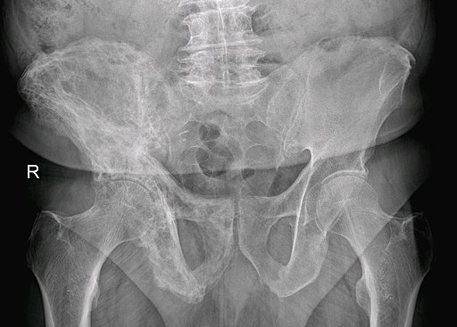
Stages and Management
| Stages | Hip (modified Ficat-Arlet) | Shoulder (Cruess) | Lunate (Lichtman) | Knee (Koshino) | Scaphoid (Herbert and Lanzetta) | Management |
| 0 – Silent | + | NWB joints – Immobilization, NSAIDs WB joints – a. Realignment osteotomy b. Core decompression +/- bone grafting or MSC therapy | ||||
| I – Suggestive clinically and MRI | + | + | + | + | + | As above NWB joints – Arthrodesis Kienbock (ulnar negative variance): Joint levelling procedure |
| II – Sclerosis or Subchondral cysts | +; IIb – Subchondral fracture (Crescent sign) | + | +; IIb – Subchondral fracture | + | + | As above |
| III – Subchondral collapse and Sphericity loss | + | + | +; IIIa – rotational dislocation of scaphoid absent; IIIb – rotational dislocation of scaphoid present (ring sign) | + | + | Arthrodesis Partial or total joint replacement Kienbock IIIb – Proximal row carpectromy |
| IV – Space narrowing and Secondary arthrosis | + | +; humeral head | + | + | + | As above |
| V – Space narrowing and Secondary arthrosis | +; also glenoid | As above |