Skip to content
Epomedicine

Mnemonics, Simplified Concepts & Thoughts

Epomedicine

Mnemonics, Simplified Concepts & Thoughts

signs of meningeal irritation

Meningeal signs

Epomedicine, Aug 18, 2014Oct 15, 2016

Meningitis refers to the inflammation of leptomeninges and underlying subarachnoid cerebrospinal fluid (CSF). Meningism or meningismus is a morbid state characterized by a meningitic syndrome (a triad of headache, photophobia and nuchal rigidity) without intracranial inflammation. Some authors, have also used the term “meningism” or “meningismus” to describe the characteristic signs of meningitis, which may be present in conditions other than meningitis. Apical pneumonia is an important cause of meningismus in children. If the meningeal signs persist for >3 days into antibiotic therapy in meningitis or the initial CSF was not fully compatible with purulent bacterial meningitis, a parameningeal suppurative focus should be considered.

Nuchal rigidity

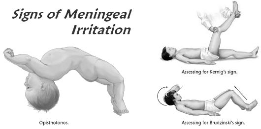
  1. Relax the neck muscles by bringing the supine patient to the edge of the bed and allow the head to hang outside the bed for a few seconds.
  2. Place your hand under the supine patient’s head and gently try to flex the neck and touch the chin to chest.
  3. Undue resistance implies diffuse irritation of cervical nerve roots from meningeal inflammation.
signs of meningeal irritation

Kernig’s sign

  1. Flex the hip and knee on one side while the patient is supine.
  2. Now, extend the knee with the hip still flexed.
  3. The sign is positive if hamstring spasm results in pain in the posterior thigh and difficulty with knee extension.
  4. With severe inflammation of meninges, the opposite knee may also flex.

Brudzinski’s neck sign (More sensitive than Kerning’s)

  1. In a supine patient, place one of your hand behind his/her head and the other on his/her chest.
  2. Now, flex the patient’s head with the hand behind the head, while your hand on chest restrains the patient and prevents him/her from rising
  3. The sign is positive if the patient flexes hips and knees.

Brudzinski’s contralateral reflex sign

  1. This has two components: the identical and reciprocal contralateral reflex.
  2. Identical contralateral reflex: On passive flexion of patient’s hip and knee on one side, the contralateral leg bends reflexly.
  3. Reciprocal contralateral reflex: This is positive when the leg contralateral leg that has flexed reflexly begins to extend spontaneously, resembling a “little kick”.

Tripod or Amoss’s or Hoyne’s sign

  1. The patient is asked to sit up in bed.
  2. Normally: sits up without supporting himself
  3. Meningeal irritation: patient tries to sit up by supporting himself with his hands placed far behind him in the bed (like a tripod), in order to take the weight off the spine and prevent its flexion. Severe meningeal irritation may result in the patient assuming the tripod position with the knees and hips flexed, the back arched lordotically, the neck extended, and the arms brought back in a plane posterior to the pelvis to support the thorax.
Meningeal Signs Elicitation Video

15 shares
  • Facebook6
  • Twitter
Clinical Skills and Approaches Clinical examinationNervous system

Post navigation

Previous post
Next post

Related Posts

Clinical Skills and Approaches

Splenomegaly : Examination techniques and Clinical Approach

Mar 20, 2021May 31, 2023

ANATOMY OF SPLEEN Histologically: FUNCTIONS OF SPLEEN Spleen is the largest lymphoid organ organ and serves following functions – An increase in these normal functions may result in splenomegaly EXAMINATION OF SPLEEN 1. Palpation: If history suggest splenomegaly but is not palpable: Roll the patient on to the right lateral…

Read More
Clinical Skills and Approaches

Ober’s Test

Oct 6, 2021Sep 5, 2022

Tensor fascial latae (TFL) originates from the anterior superior iliac spine (ASIS) outer surface, outer lip of anterior iliac crest and deep surface of fascia latae, continues down in thigh as Iliotibial band (ITB) and inserts to the ipsilateral Gerdy’s tubercle. TFL plays role in flexion, medial rotation and abduction…

Read More
Clinical Skills and Approaches

CNS examination long case: How to prepare

Jul 8, 2020Jul 8, 2020

Central nervous system (CNS) examination is the most difficult thing to complete within a time limit during final exam and the dilemma is that, most of time the CNS is what is kept as a long case. Examiners love judging you on the basis of CNS case because: 1. CNS…

Read More

Leave a Reply Cancel reply

Your email address will not be published. Required fields are marked *

This site uses Akismet to reduce spam. Learn how your comment data is processed.

Epomedicine. Meningeal signs [Internet]. Epomedicine; 2014 Aug 18 [cited 2026 Jul 28]. Available from: https://epomedicine.com/clinical-medicine/cns-examination-meningeal-signs/.

Pre-clinical (Basic Sciences)

Anatomy

Biochemistry

Community medicine (PSM)

Embryology

Microbiology

Pathology

Pharmacology

Physiology

Clinical Sciences

Anesthesia

Dermatology

Emergency medicine

Forensic

Internal medicine

Gynecology & Obstetrics

Oncology

Ophthalmology

Orthopedics

Otorhinolaryngology (ENT)

Pediatrics

Psychiatry

Radiology

Surgery

RSS Ask Epomedicine

  • What to study for Clinical examination in Orthopedics?
  • What is the mechanism of AVNRT?

Epomedicine weekly

  • About Epomedicine
  • Contact Us
  • Author Guidelines
  • Submit Article
  • Editorial Board
  • USMLE
  • MRCS
  • Thesis
©2026 Epomedicine | WordPress Theme by SuperbThemes