Skip to content
Epomedicine

Mnemonics, Simplified Concepts & Thoughts

Epomedicine

Mnemonics, Simplified Concepts & Thoughts

Clarke’s test

Epomedicine, Aug 12, 2023Aug 12, 2023

Synonyms: Patellofemoral grind test, Patella hold test, Zohler’s sign

Tests for: Patellofemoral dysfunction

Patient position: Lying down with knees extended and relaxed (heels on table)

Technique:

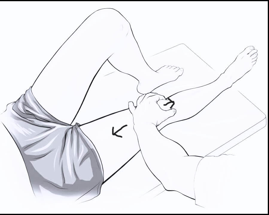
1. Place the webspace of thumb on the upper pole of patella and push it inferiorly.

2. Ask the patient to contract his/her quadriceps muscle.

clarke's test

Interpretation:

  1. Positive test: Retro-patellar pain and patient cannot hold the contraction
  2. Negative test: No pain and patient can hold the contraction

2 school of thoughts:

1st thought: The examiner can achieve a positive test on anyone if sufficient pressure is applied to the patella. Hence, the test must be repeated several times, increasing the pressure each time and comparing the results with those of the unaffected side. To test different parts of the patella, the knee should be tested in 30, 60 and 90 degrees of flexion besides full extension.1Magee, D. J. (2008). Orthopedic Physical Assessment. Malawi: Saunders Elsevier.

2nd thought: Since, it is a painful test, it should be performed only once and at the end of the examination.2The Art of the Musculoskeletal Physical Exam. (2023). Switzerland: Springer International Publishing.

  • Facebook
  • Twitter
Clinical Skills and Approaches Clinical examinationMusculoskeletal systemOrthopedics

Post navigation

Previous post
Next post

Related Posts

Clinical Skills and Approaches

C-spine X-ray : Mnemonic Approach

Oct 27, 2020Sep 22, 2022

Mnemonic: ABCDEF For images of the particular Cervical spine (C-spine) X-ray findings and views mentioned below, please refer to the links at the end of the article or use web-search. Adequacy Skull base, C1-C7 and upper T1 must be visible 3 views: True lateral, AP, Odontoid (Open mouth) +/- Swimmer’s…

Read More
Clinical Skills and Approaches complications of transfusion

Perioperative Fluid Management

Jan 1, 2016May 5, 2016

Author: Sulabh Kumar Shrestha, KISTMCTH A) RELEVANT ANATOMY AND PHYSIOLOGY Details of the body fluid compartments are described here Microvessels for fluid exchange: The Exchange Vessels – capillaries and most proximal part of the venules Sinusoidal capillaries (liver, spleen, bone marrow): freely permeable to all solutes Fenestrated capillaries (glands, glomeruli, GIT):…

Read More
Clinical Skills and Approaches history and examination format

Cyanosis

Dec 3, 2013Feb 16, 2018

Synonyms: Morbus coeruleus, Blue disease Definition of Cyanosis Cyanosis is the bluish discoloration of the skin, mucous membrane and nail bed usually owing to atleast 5 gm/dl of reduced hemoglobin/deoxyhemoglobin or abnormal hemoglobin derivatives (eg. methemoglobin, sulfhemoglobin, etc.) circulating within the superficial dermal capillaries and subpapillary venous plexus (not, as commonly taught, the…

Read More

Leave a Reply Cancel reply

Your email address will not be published. Required fields are marked *

This site uses Akismet to reduce spam. Learn how your comment data is processed.

Pre-clinical (Basic Sciences)

Anatomy

Biochemistry

Community medicine (PSM)

Embryology

Microbiology

Pathology

Pharmacology

Physiology

Clinical Sciences

Anesthesia

Dermatology

Emergency medicine

Forensic

Internal medicine

Gynecology & Obstetrics

Oncology

Ophthalmology

Orthopedics

Otorhinolaryngology (ENT)

Pediatrics

Psychiatry

Radiology

Surgery

RSS Ask Epomedicine

  • What to study for Clinical examination in Orthopedics?
  • What is the mechanism of AVNRT?

Epomedicine weekly

  • About Epomedicine
  • Contact Us
  • Author Guidelines
  • Submit Article
  • Editorial Board
  • USMLE
  • MRCS
  • Thesis
©2026 Epomedicine | WordPress Theme by SuperbThemes