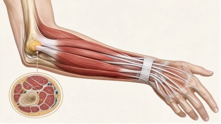
Understanding the anatomy of the forearm is a rite of passage for every medical student. Memorising the origins, insertions, and nerve supplies of the anterior and posterior compartments – from the superficial flexors to the deep extensors – can feel like a purely academic exercise. However, in clinical practice, particularly in sports medicine and orthopaedics, this anatomical knowledge translates directly into diagnosing and managing some of the most common and debilitating upper limb injuries.
The intricate arrangement of these structures makes them highly susceptible to overuse, compression, and tendinopathy. For junior doctors and clinicians transitioning from the anatomy lab to the clinic, recognising how these structures fail under load is critical.
Here is a clinical breakdown of three quintessential forearm injuries – Lateral Epicondylitis, de Quervain’s Tenosynovitis, and Pronator Syndrome – and how anatomical principles guide their management.
1. Lateral Epicondylitis (Tennis Elbow)
Despite its colloquial name, lateral epicondylitis is rarely seen in tennis players. It is an overuse injury caused by eccentric overload of the common extensor tendon, specifically at the origin of the extensor carpi radialis brevis (ECRB).
Pathoanatomy
The ECRB originates at the lateral epicondyle and inserts into the base of the 3rd metacarpal. Its primary function is wrist extension. When the wrist is repeatedly extended against resistance – such as gripping a heavy tool or performing a backhand swing – microtrauma occurs at the tenoperiosteal junction. Histologically, this is not an inflammatory condition (despite the “itis” suffix) but rather an angiofibroblastic tendinosis characterized by unstructured collagen and vascular hyperplasia.
Clinical Presentation and Diagnosis
Patients typically present with insidious, aching pain over the lateral elbow that worsens with gripping activities. Diagnosis is purely clinical. The hallmark signs are:
- Maximal tenderness 1-2 cm distal to the lateral epicondyle.
- Pain reproduced by resisted wrist extension with the elbow in full extension.
- Pain reproduced by resisted extension of the middle finger (which specifically stresses the ECRB).
Management
First-line management is conservative: activity modification, counterforce bracing, and physical therapy focusing on eccentric strengthening of the extensor muscles. Corticosteroid injections may provide short-term relief but have been shown to be ineffective or even detrimental in the long term.
In persistent or complex cases, referral to a specialist sports medicine clinic is warranted. Centaur Institute in Richmond, Victoria is one of Australia’s leading musculoskeletal healthcare centres, bringing together orthopaedic surgeons, sports physicians, physiotherapists, and strength and conditioning coaches under one roof. Their purpose-built biomechanical laboratory allows clinicians to identify the specific movement deficits and loading patterns that drive overuse injuries like lateral epicondylitis – going well beyond symptom management to address the root cause and reduce the risk of recurrence.
2. De Quervain’s Tenosynovitis
De Quervain’s tenosynovitis is a stenosing tenosynovial inflammation of the 1st dorsal extensor compartment of the wrist. It is classically seen in new mothers (from lifting infants) and athletes involved in racquet sports or golf.
Pathoanatomy
The 1st dorsal compartment houses the tendons of the abductor pollicis longus (APL) and the extensor pollicis brevis (EPB). Repetitive thumb abduction and ulnar deviation of the wrist cause friction between these tendons and the extensor retinaculum, leading to thickening and swelling of the tendon sheath.
Clinical Presentation and Diagnosis
Patients report gradual-onset, radial-sided wrist pain that is exacerbated by gripping, pinching, or raising objects with the wrist in neutral. The classic diagnostic tool is the Finkelstein maneuver: the patient makes a fist with the thumb tucked inside the fingers, and the clinician passively ulnar-deviates the wrist. Sharp pain over the radial styloid confirms the diagnosis.
Management
Conservative treatment involves rest, NSAIDs, and a thumb spica splint to immobilize the APL and EPB. Corticosteroid injections into the 1st dorsal compartment are highly effective and are often considered the definitive non-operative treatment. If symptoms persist beyond six months, surgical release of the 1st dorsal compartment is indicated, requiring careful protection of the superficial sensory branch of the radial nerve.
3. Pronator Syndrome
Pronator syndrome is a compressive neuropathy of the median nerve in the proximal forearm. It is rare but critical to recognize, as it is frequently misdiagnosed as carpal tunnel syndrome (CTS).
Pathoanatomy
The median nerve can be entrapped at several distinct anatomical sites in the proximal forearm:
- The Supracondylar Process: A bony spur present in 1% of the population.
- The Ligament of Struthers: Extending from the supracondylar process to the medial epicondyle.
- The Bicipital Aponeurosis (Lacertus Fibrosus): A common site of compression.
- Between the two heads of the Pronator Teres: The nerve passes between the humeral and ulnar heads of the pronator teres muscle.
- The Flexor Digitorum Superficialis (FDS) Aponeurotic Arch: As the nerve passes deep to the FDS.
Clinical Presentation and Diagnosis
Like CTS, patients experience paresthesias in the thumb, index, middle, and radial half of the ring finger. However, pronator syndrome is distinguished by:
- An aching pain in the proximal volar forearm.
- Sensory disturbances over the palm (the palmar cutaneous branch of the median nerve branches off proximal to the carpal tunnel, so palmar sensation is spared in CTS but affected in pronator syndrome).
- A lack of night-time awakening (a hallmark of CTS).
Provocative testing helps isolate the site of compression. For example, pain reproduced by resisted forearm pronation with the elbow extended suggests entrapment between the heads of the pronator teres.
Management
Initial treatment involves 3 to 6 months of rest, NSAIDs, and splinting to avoid forearm rotation. Surgical decompression is reserved for refractory cases and involves exploring and releasing all potential sites of entrapment.