The earliest trial-enrollment window is often the point when a parkinsonian diagnosis is least settled. Parkinson’s disease, progressive supranuclear palsy (PSP), and multiple system atrophy (MSA) can initially share bradykinesia, rigidity, and some response to dopaminergic therapy before distinguishing features become clear.
The differential diagnosis of atypical parkinsonian syndromes therefore poses a timing problem for clinical research. Investigators need credible cohort definitions before longitudinal observation resolves the uncertainty, particularly when disease-modifying therapies target different underlying pathologies.
Quantitative structural MRI and molecular imaging approach that uncertainty from different directions. Their research value depends on understanding which biological question each measurement addresses, where their findings overlap, and what remains unproven when early imaging appears to support a particular diagnosis.
The Short Answer: MRI Narrows, Molecular Imaging Confirms
Quantitative MRI and molecular imaging aren’t competing answers to the same question. MRI localizes structural change; molecular imaging characterizes selected dopaminergic and metabolic processes. Together, they can strengthen the differential diagnosis of atypical parkinsonian syndromes without replacing clinical assessment or establishing protein pathology.
The limitation of transporter imaging is diagnostic overlap. Parkinson’s disease, PSP, MSA, and corticobasal degeneration (CBD) can all produce reduced binding. An abnormal scan therefore doesn’t identify which disorder caused the deficit.
Structural measurements likewise support a PSP profile; they don’t establish pathology. Parkinson’s disease typically lacks a specific conventional brainstem atrophy pattern, but a normal midbrain-to-pons ratio doesn’t prove that diagnosis or exclude early PSP. Thresholds also require validation in the phenotype and acquisition setting being studied.
Reading the Brainstem: What Quantitative MRI Measures
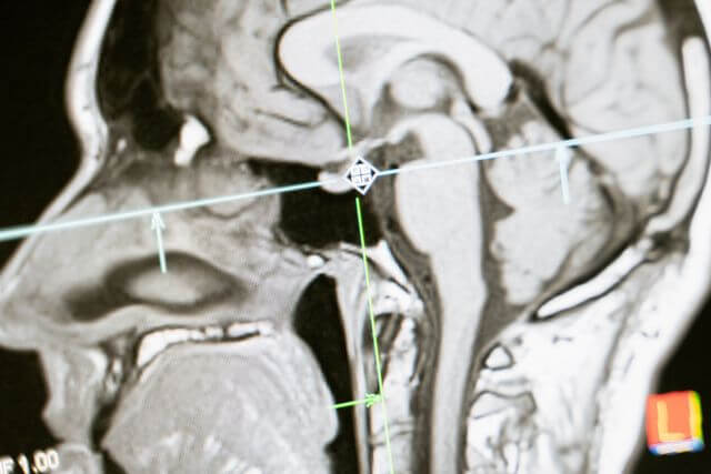
Visual magnetic resonance imaging (MRI) signs, including the “hummingbird” appearance associated with PSP, aren’t consistently apparent early. Quantitative volumetrics and morphometry turn subjective brainstem impressions into reproducible imaging biomarkers that can support comparisons across scanners, sites, and visits when acquisition, processing, and quality control are standardized.
Midbrain-to-Pons Ratio and the MR Parkinsonism Index
PSP-Richardson’s syndrome characteristically involves disproportionate midbrain atrophy relative to the pons. The midbrain-to-pons area ratio expresses that anatomical imbalance: a smaller midbrain relative to the pontine reference produces a lower ratio.
Importantly, this is an area-based measurement, not a volumetric measurement, although both belong within quantitative structural assessment.
The MR Parkinsonism Index combines the inverse brainstem relationship, pons-to-midbrain area, with the middle-to-superior cerebellar peduncle width ratio. This incorporates another anatomical feature relevant to PSP rather than relying on one structure alone.
The study “Diagnostic Potential of Multimodal MRI Markers in Atypical Parkinsonian Disorders” examines the diagnostic contribution of complementary MRI markers, supporting assessment beyond a single visual sign.
Pontine and Putaminal Signatures of MSA
MSA requires a different anatomical reading. Pontine and middle cerebellar peduncle atrophy contribute to its structural profile, while putaminal volume loss is particularly relevant to MSA-P, the parkinsonian subtype. Their prominence varies with phenotype and disease stage.
Because pontine shrinkage changes the denominator, the midbrain-to-pons ratio can remain relatively preserved or move upward rather than follow the PSP pattern. Treating that ratio as a universal atypical-parkinsonism score would therefore misread its purpose.
Automated segmentation reduces manual measurement burden but doesn’t eliminate error. Multicenter studies still need inspection of anatomical boundaries, consistent image acquisition, and control of scanner-related variation before interpreting longitudinal volume change.
Where DAT-SPECT Stops and PET Picks Up the Signal
“Molecular imaging” doesn’t describe a single diagnostic test. Presynaptic dopamine transporter imaging, postsynaptic receptor imaging, and metabolic PET imaging measure different processes. An abnormal result only answers the biological question that its tracer was designed to investigate.
Why DAT Loss Appears in Every Degenerative Parkinsonism
DAT-SPECT assesses dopamine transporter availability in the striatum. Reduced striatal binding supports presynaptic nigrostriatal dysfunction and helps distinguish degenerative parkinsonism from conditions without that deficit, including essential tremor and uncomplicated drug-induced parkinsonism. Medication effects and scan quality still require review.
As noted above, laterality and caudate-to-putamen gradients can contribute to interpretation, but group-level patterns aren’t reliable individual disease labels. A participant with asymmetric transporter loss doesn’t automatically belong in a Parkinson’s disease cohort, particularly when clinical or structural findings suggest an atypical syndrome.
What 18F-DMFP and Metabolic PET Add to the Picture
18F-DMFP PET evaluates postsynaptic D2/D3 receptor availability. Relative preservation in Parkinson’s disease contrasts with reductions associated with striatal involvement in PSP and MSA. Nevertheless, receptor availability isn’t a direct neuronal count: treatment exposure and biological regulation complicate interpretation.
Metabolic PET adds a regional functional perspective, assessing distributions of reduced metabolism rather than transporter loss alone. Disease-associated patterns can strengthen a differential diagnosis when they agree with clinical and structural findings.
Support vector machine and Bayesian network classifiers provide ways to combine these distributed PET signals into multiclass predictions. Their apparent discrimination isn’t sufficient evidence of clinical readiness, however.
Training-cohort composition, independent validation, and the reliability of diagnostic labels determine whether a classifier generalizes beyond its development dataset.
Combining Modalities to Build Cleaner Trial Cohorts
The useful combination is complementary evidence, not a tally of positive scans. Structural measurements characterize anatomical injury, while molecular measurements characterize dopaminergic or metabolic dysfunction. Agreement strengthens a diagnostic profile; disagreement identifies uncertainty that shouldn’t disappear inside an enrollment label.
Stacking Structural and Molecular Markers Early
A research protocol can use abnormal DAT-SPECT to support nigrostriatal dysfunction, then assess whether brainstem and putaminal measurements favor PSP, MSA-P, or neither. Postsynaptic or metabolic PET can address residual ambiguity.
This is a protocol-dependent sequence, not a requirement that every participant undergo every modality.
Structural MRI is generally easier to repeat and more accessible than specialized PET. Availability of 18F-DMFP, tracer production, and imaging costs constrain its use, so selective PET assessment is more practical than assuming universal access.
Combined interpretation also requires predefined rules. Centralized quantitative reads, standardized acquisition, and documented quality-control failures prevent site differences from masquerading as biological differences.
Protocols should specify how discordant findings affect eligibility rather than resolving them retrospectively after treatment outcomes become available.
Tauopathy or Synucleinopathy: Matching Patients to Targets
For tau-targeted disease-modifying therapies, a PSP-compatible clinical and imaging profile provides stronger enrollment support than parkinsonism alone. Conversely, an MSA-compatible profile supports investigation within an α-synucleinopathy framework.
Neither structural MRI nor dopaminergic imaging directly proves the responsible protein pathology.
Corticobasal syndrome (CBS) needs particular care: it is a clinical syndrome, not a synonym for CBD or a guaranteed tauopathy. A tau-directed study shouldn’t classify every CBS participant as biologically eligible solely from the syndrome label.
Misclassification reduces the proportion of participants whose disease matches the treatment target. That can dilute an observed treatment effect and weaken statistical power, even when the intervention acts in the intended subgroup.
Baseline volumetric measurements can also become longitudinal research endpoints. Their use as progression measures requires separate validation; a marker that distinguishes diagnoses isn’t automatically sensitive to change or established as a surrogate for treatment benefit.
What This Means for Early Parkinsonism Research
For early-stage trials, the defensible approach is a predefined, validated combination of evidence, with explicit handling of uncertainty. Disease-modifying research depends on diagnostic specificity at enrollment, not simply on a diagnosis that becomes convincing years later.
Complementary imaging supports that goal when its limitations remain visible in cohort definitions and endpoint selection.