Skip to content
Epomedicine

Mnemonics, Simplified Concepts & Thoughts

Epomedicine

Mnemonics, Simplified Concepts & Thoughts

rubeosis iridis neovascular glaucoma

Ophthalmology spot diagnosis : Rubeosis Iridis

Epomedicine, May 23, 2014Jul 2, 2014


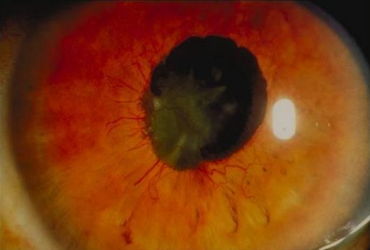
Definition: Neovascularization of iris

Pathophysiology: Causes that lead to retinal hypoxia triggers release of vasoproliferative factors include vascular endothelial growth factor (VEGF), fibroblast growth factor (FGF) and others

rubeosis iridis neovascular glaucoma

Etiology:

1. Diabetic Retinopathy

2. Retinal Vascular Occlusive Diseases

  • Central Retinal Vein Occlusion (CRVO)
  • Ischaemic Hemiretinal Vein Occlusion

3. Ocular Ischaemic Syndrome

  • Carotid Artery Occlusive Disease
  • Takayasu’s Syndrome
  • Carotid-Cavernous Fistula
  • Giant Cell Arteritis
  • Wyburn-Mason syndrome
  • Strabismus Surgery
  • Ocular Radiation

4. Tumours

  • Uveal Melanomas
  • Metastatic Choroidal Tumours
  • Medulloepithelioma
  • Hypoxic Retinoblastoma
  • Pigmented Ciliary Adenocarcinoma
  • Metastatic Malignant Lymphoma

5. Others

  • Uveitis
  • Retinal Vasculitis
  • Coat’s Disease (Retinal telangiectasia)
  • Eales’ Disease (Periphlebitis retinae)
  • Sarcoidosis
  • X-linked Retinoschisis
  • Chronic Retinal Detachment
  • Retinopathy of Prematurity (ROP)
  • Systemic Cryoglobulinaemia

Important causes:

  1. Proliferative Diabetic Retinopathy (PDR)
  2. Central Retinal Vein Occlusion (CRVO)
  3. Sickle cell retinopathy
  4. Chronic iridocyclitis
  5. Retinoblastoma

Grading of rubeosis iridis:

  • 0 : No iris neovascularization
  • 1 : Less than 2 quadrants of NV at iris pupillary zone
  • 2 : More than 2 quadrants of NV at iris pupillary zone
  • 3 : Grade 2 + less than 3 quadrants of NV at iris ciliary zone and/or ectropion uveae
  • 4 : More than 3 quadrants of NV at iris ciliary zone and/or ectropion uveae

Findings:

  1. Abnormal iris vessels
  2. Perform gonioscopy to assess presence of angle neovascularization
  3. May have elevated IOP (neovascular glaucoma)

Rubeosis Iridis and Neovascular glaucoma:

The disease develops in 3 stages:

1. Neovascularization of the iris (NVI)

2. Secondary open angle glaucoma (SOAG): The NVI extend to involve the angle, and are accompanied by fibrosis, blocking the trabecular meshwork and  causing ocular hypertension, and SOAG.

3. Secondary angle closure glaucoma (SACG): Myofibroblasts within the fibrovascular tissue proliferate and contract, forming peripheral anterior synechiae (PAS), and secondary angle closure, with resulting intra-ocular pressure rise.

100 days glaucoma: Neovascular glaucoma (NVG) secondary to ischemic CRVO

Treatment:

  1. Ischemia: Panretinal Photocoagulation (PRP)
  2. IOP control: Glaucoma drainage implant (e.g. Molteno’s tube)
41 shares
  • Facebook18
  • Twitter
Case Reports

Post navigation

Previous post
Next post

Related Posts

Case Reports

Case of Cyanotic Congenital Heart Disease : PGE1 saves life

Jun 2, 2020Jun 2, 2020

A Single Male baby was born at 38 weeks of gestation with birth weight of 3.1 kg through Normal vaginal delivery. At birth the child had cried immediately. At 15 minutes of life, the child developed central cyanosis. There was no respiratory distress and heart rate was normal range. Child…

Read More

A Case of Neuroleptic Malignant Syndrome: A Quick Approach

Dec 6, 2013Jun 12, 2016

A 15 year girl started complaining of strange feelings 3 weeks back. She started behaving weirdly and complained her mother that she could hear inanimate objects talking to her- like bangles, rings, glasses. She rapidly had a deterioration with change in personality. She starting acting bizarre and doing abnormal things….

Read More
Case Reports Jones criteria

A Case of Rheumatic Fever

Jan 3, 2014Jul 14, 2020

A 12 year old female came with complaints of multiple joint pain for 10 days which started in the left knee and resolved within 3 days, then started on the bilateral ankles which also resolved in 3 to 4 days and started on the right knee. She currently complains of…

Read More

Comment

  1. Mustafa Shuaib says:
    May 11, 2016 at 6:05 pm

    Good, it’s nice note, i really get new information about this topic

    Reply

Leave a Reply to Mustafa Shuaib Cancel reply

Your email address will not be published. Required fields are marked *

This site uses Akismet to reduce spam. Learn how your comment data is processed.

Epomedicine. Ophthalmology spot diagnosis : Rubeosis Iridis [Internet]. Epomedicine; 2014 May 23 [cited 2025 Dec 30]. Available from: https://epomedicine.com/clinical-cases/ophthalmology-spot-diagnosis-rubeosis-iridis/.

Pre-clinical (Basic Sciences)

Anatomy

Biochemistry

Community medicine (PSM)

Embryology

Microbiology

Pathology

Pharmacology

Physiology

Clinical Sciences

Anesthesia

Dermatology

Emergency medicine

Forensic

Internal medicine

Gynecology & Obstetrics

Oncology

Ophthalmology

Orthopedics

Otorhinolaryngology (ENT)

Pediatrics

Psychiatry

Radiology

Surgery

RSS Ask Epomedicine

  • What to study for Clinical examination in Orthopedics?
  • What is the mechanism of AVNRT?

Epomedicine weekly

  • About Epomedicine
  • Contact Us
  • Author Guidelines
  • Submit Article
  • Editorial Board
  • USMLE
  • MRCS
  • Thesis
©2025 Epomedicine . All rights reserved.