


Definition of Apoptosis
Apoptosis is:
- Energy (ATP) – dependent, genetically Programmed Cell Death (PCD) in which,
- Cells activate enzymes that degrade the cells’ own nuclear DNA and nuclear cytoplasmic protein.
Origin of the word “apoptosis”:
- Derived from Greek word
- Apo = separation + Ptosis = falling off (especially used for leaves of flower petals)
Caspases – Central Regulators of Apoptosis
Caspases = Cysteine Aspartate Specific Proteases
2 types of caspases:
Initiator caspases:
- Caspase-2, -8, -9, -10, -11, and -12
- Initiator of extrinsic pathway: Caspase-8 and -10
- Initiator of intrinsic pathway: Caspase-2 and -9
- Closely coupled to pro-apoptotic signals
- Once, activated – initiator caspases cleaves and activates effector or executioner caspases 1http://www.cellsignal.com/contents/science-pathway-research-apoptosis/regulation-of-apoptosis-signaling-pathway/pathways-apoptosis-regulation
Executioner caspases:
- Caspase -3, -6, and -7
- Execute apoptosis in 2 ways:
- Itself is a protease: cleaves cytoskeletal proteins
- Activates endonuclease (eg. CAD – Caspase Activated DNAse): cleaves DNA and nucleoproteins
3 Pathways of Caspase Activation
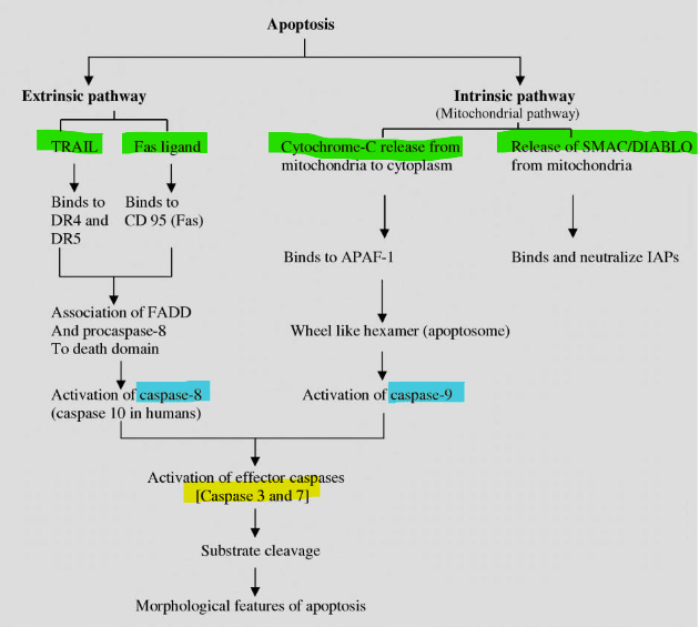
1. Activation of Initiator of Extrinsic Pathway
Receptor-ligand interaction mediated:
- FAS receptor (FASR or CD95) activation on target cell by FAS Lignad expressed by activated T lymphocytes
- Type I TNF receptor activation on target cell by TNF
Can mediate intrinsic (mitochondrial) pathway as well:
- Caspase-8 may cleave and activate a pro-apoptotic member of the Bcl-2 family called Bid which mediates mitochondrial pathway
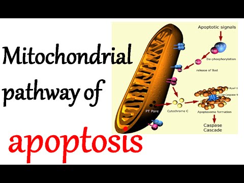
2. Activation of Initiator of Intrinsic Pathway
DNA damage or Loss of hormonal stimulation leads to:
- Activation of Caspase-2 mediated by PIDDosome which forms Caspase-2 activation complex
- Caspase-2 activation complex is comprised of:
- Caspase-2
- PIDD (p-53 inducible death domain)
- RAIDD (RIP-associated Ich-1/Ced-3-homologue protein with a death domain)

Activation of caspase-2 leads to inactivation of anti-apoptotic BCL-2 protein and activation of pro-apoptotic BAX, BAK and BID which insert into mitochondrial membrane
Cytochrome C into the cytosol from mitochondria through the channels formed by the pro-apoptotic proteins
Cytochrome C binds with Apaf-1 (apoptotic protease activating factor-1) protein in cytosol to form Apoptosome
Caspase-9 binds to Apoptosome and gets activated
Along with cytochrome C, other proteins that leak from mitochondrium are:
- Inhibitor of Apoptosis (IAP): inhibits executioner caspase (-3, -7), and initiator caspase (-9) by inhibiting caspase catalytic activity and ubiquitin-proteasome cleavage of caspases.
- Pro-apoptotic: Smac/Diablo, AIF, HtrA2, and Endo G.; Smac/Diablo binds inhibits caspase inhibitor XIAP
3. Perforin/Granzyme pathway
A pathway utilized by cytotoxic T cells
Perforins secreted by CD8+ T cell create pores in target cell membranes
Granzyme from CD8+ T cell enters pores:
- Granzyme B:
- Activates mitochondrial pathway
- Direct activation of caspase-3
- Granzyme A:
- Activation of a type of DNAse 2Apoptosis: A Review of Programmed Cell Death – Susan Elmore
Endpoint of Apoptosis – Clearance of Apoptotic cells
Phagocytosis of apoptotic cells occur before it leaks out and mediate inflammation, primarily by 2 mechanisms:
- Inner phospholipid layer of plasma membrane “flips” in apoptotics cells – recognized by macrophages
- Release of soluble factors that recruit phagocytes
For a greater detail – view the apoptosis map in humans.
Anti-apoptotic ligands, including growth factors and cytokines, activate Akt and p90RSK. Akt inhibits Bad by direct phosphorylation and prevents the expression of Bim by phosphorylating and inhibiting the Forkhead family of transcription factors (FoxO). FoxO promotes apoptosis by upregulating pro-apoptotic molecules such as FasL and Bim.
Morphology or Histological Changes of Apoptosis
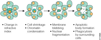
- Cell shrinkage and eosinophilic cytoplasm
- Pyknosis (Nuclear condensation) and Karyorrhexis (Nuclear fragmentation)
- Membrane blebbing and formation of apoptotic bodies
- Phagocytosis of apoptotic bodies
Examples of Apoptosis
Physiologic
- During embryogenesis
- Endometrial shedding during menstruation
- Inflammatory cells after serving the purpose
- Negative selection of self-reactive lymphocytes
Pathologic
- Failed repair of DNA damage with probability of mutation
- Accumulation of misfolded proteins (ER stress leading to Caspase-12 activation), eg. Alzheimer’s disease, Parkinson’s disease, Type 2 Diabetes Mellitus
- HIV or Hepatitis infection
- Gland atrophy following duct obstruction, eg. pancreas, parotid 3Robbin’s Basic Pathology – 9th edition
Diagnosis of Apoptosis
1. Chromatin condensation seen by hematoxylin, Feulgen and acridine orange staining.
2. Estimation of cytochrome ‘c’
3. Estimation of activated caspase
4. Estimation of Annexin V (apoptotic cells express phosphatidylserine on the outer layer of plasma membrane because of which these cells are recognized by the dye Annexin V. Some cells also express high concentration of thrombospondin).
5. DNA breakdown at specific sites can be detected by ‘step ladder pattern’ on gel electrophoresis or TUNEL (TdT mediated d-UTP Nick End Labelling) technique.
Clinical Relevance – Apoptosis and Diseases
Decreased Apoptosis
- Autoimmune diseases
- Due to failed negative selection of auto-reactive lymphocytes.
- Potent inducers of apoptosis like steroids and cyclophosphamide are effective in autoimmune diseases.
- Hematological malignancies
- Increase in Fas/CD95 expression has been found on myeloid progenitor cells from patients with chronic myeloid leukemia
(CML), myelodysplasia and aplastic anemia compared to normal marrow progenitors. - Bcl-2 expression correlates with a poor response to chemotherapy in acute myeloid leukemia.
- Expression of Bcl-2 is down regulated after treatment with a signal transduction inhibitor (Imatinib) – suggesting involvement in CML.
- Deletions or mutations of p53 have been noted in many hematological disorders including: CML and CLL.
- Increase in Fas/CD95 expression has been found on myeloid progenitor cells from patients with chronic myeloid leukemia
- Viral infections
- Both p35 gene and inhibitor of apoptosis (IAP) found in baculovirus are able to inhibit apoptosis in a response to large number of triggers.
- Pox virus inhibit apoptosis by producing IL-1 converting Enzyme (ICE)
Increased Apoptosis
- AIDS
- Induction of CD4+ T cell apoptosis
- Neurodegenerative diseases
- In acute neurological diseases, both necrosis and caspase-mediated apoptotic cell death occur.
- By contrast, in chronic neurodegenerative diseases, caspase-mediated apoptotic pathways have the dominant role.
- Beta-amyloid proteins that accumulate in Alzheimer’s disease induces neuronal apoptosis
- Minocycline (apoptosis inhibitor) has been on trial for Huntington’s disease and ALS 4Apoptosis and Diseases – Relevance and Clinical Significance by Faris Q. Alenzi, PhD, FAMLI