
Etiology
Mnemonic: RSTUV
- Radial inclination – decreased
- Shape of lunate (Type 1 has more proximal apex; Type 2 & 3 are more rectangular)
- Type 1 lunate is seen with negative ulnar variance and possess highest risk of Kienbock’s disease
- Trauma (repetitive micro-fractures or single fracture)
- Ulnar variance – negative (increased radial-lunate contact stress)
- Vascular anatomy (3 patterns – X, Y, I)
- “I” pattern (single vessel to lunate) – highest risk of avascular necrosis
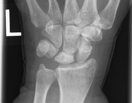
Muzichick, CC BY-SA 4.0, via Wikimedia Commons
Lichtman Classification and Management
| Stage | Description | Treatment |
| Mnemonic: ABCD | Mnemonic: ABCD | |
| I | Abnormal MRI (decreased T1 intensity; variable T2 intensity) or scintigraphy | Analgesics + immobilization |
| II | Bone sclerosis ± Bone breaks (fracture lines) | Bony procedures: 1. Negative or Neutral ulnar variance: Joint levelling procedure (Radius shortening osteotomy; Ulnar lengthening) 2. Positive ulnar variance: Revascularization procedures (pedicled vascularized bone graft from dorsal distal radius), Distal radius core decompression, Radial wedge osteotomy |
| III | Collapse of wrist with: | |
| A | Normal carpal alignment | Same as stage II |
| B | Fixed scaphoid rotation | Carpal fusion (STT or SC) Carpectomy (PRC) |
| IV | Degenerative changes of wrist | Deliverance (Salvage) 1. Proximal row carpectomy (PRC – allows capitate to articulate into lunate fossa) 2. Wrist arthrodesis 3. Wrist denervation 4. Total wrist arthroplasty |


He is the section editor of Orthopedics in Epomedicine. He searches for and share simpler ways to make complicated medical topics simple. He also loves writing poetry, listening and playing music.