
Following events occur in the zygote formed in the week zero of development:
Cleavage
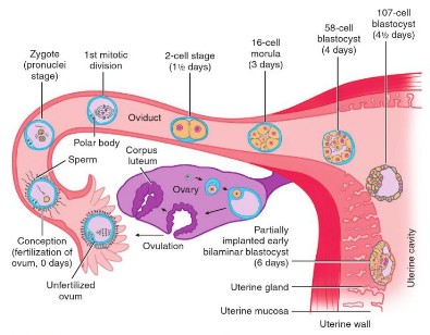
- Approximately 24 hours after fertilization the zygote begins with the first cleavage division.
- Series of mitotic divisions of the zygote (occurs in fallopian tube) to form small daughter cells called blastomeres.
- Characteristics of cleavage in humans:
- Holoblastic: Complete cleavage that divides the whole egg into distinct and separate blastomeres due to absence of yolk. In hen’s egg the cleavage is incomplete (meroblastic) due to presence of large amount of yolk.
- Asymmetrical: Blastomeres are unequal in size (unequal cytoplasm distribution)
- Asynchronous: Occuring at different times
- Each cleavage takes around 12 to 24 hours to complete.
Morula vs Blastula vs Blastocyst
I know, most of you are confused about these terms because different authors have mentioned these terms with different meanings. Let’s make these terms clear:
Morula: After completion of several cleavage divisions, the blastomeres are arranged in the form of a solid of cells like mulberry by a process called compaction. This mulberry like structure is morula.
- Consists of 16 blastomeres
- Formed around day 3
- It has an inner and outer cell mass
Blastula: Rearrangement of blastomeres to form a hollow spherical ball with an inner cavity called Blastocoel is called Blastula.
- Consists of around 128 blastomeres surrounding the blastocoel.
- It is present in non-mammalians.
Blastocyst: In simple words, blastula in mammals is called Blastocyst. However, the cells of blastula and blastocyst have different fates.
- It is present in mammalians.
- Formed around day 4-5.
- Outer cell mass forms trophoblast and Inner cell mass forms embryoblast.
- It is not the morula, but the blastocyst that attaches itself to the endometrium of uterus.
While the cells of blastula give rise to all the structures of adult organism in future, only the embryoblast (inner cell mass) of blastocyst gives rise to all the structures of adult organism in future and the outer trophoblast (outer cell mass) forms placenta which is discarded at birth. So, the basic difference is that the non-mammalian blastula comprises of single functional layer and mammalian blastula consists of 2 different functional cell layers. 1Understanding Human Anatomy Through Evolution – Second Edition By Bruce D. Olsen
How is blastocoel formed ?
- The trophoblasts have Na+ pumps and exchangers that pump Na+ into centrally forming cavity.
- This creates an osmotic drive that pulls the water inside the cavity.
Transport of Zygote
Zygote ascends in the fallopian tube and towards uterine cavity aided by 2 mechanisms:
- Tubal cilia transport
- Tubal muscle contraction
Implantation
On around the 6th day of development, after the zona pellucida has disappeared, the trophoblast at the embryonic pole loosely attaches to the functional layer of endometrium (endometrium is in secretory phase during this time).
Site: Posterior superior wall of uterus
Mechanism: Similar to inflammation –
- L selectin on trophoblast cells and its carbohydrate receptors on the uterine epithelium mediate initial attachment of the blastocyst to the uterus.
- Integrins mediate invasion and migration.
| Day | 0 | 1 | 2 | 3 | 4 | 5 | 6 |
| Cell number | 1 | 2 | 4-8 | 16 | 32 | 128 | |
| Events | Fertilization | First cleavage | Continuing cleavage | Morula | Early blastocyst | Late blastocyst | Implantation starts |
Ectopic Pregnancy
A defect in transport of zygote (ciliary or tubal muscle) and it’s implantation outside the uterine cavity, along the fallopian tube leads to a condition called Ectopic pregnancy. In such cases, amnion and chorion are formed but a true decidua is never present.
Dizygotic (Fraternal) Twinning
As the name suggests, “Dizygotic” twins develop from 2 different zygotes originating from fertilization of 2 oocytes each by a different sperm. Hence, the dizygotic twins:
- Are different genetically
- Can have same or different sex
- Have 2 separate chorion, amnion and amnion (dichorionic and diamniotic) – the zygotes implant individually in the uterus and proceed with development.
- However, due to close apposition – 2 placentas or chorionic sacs may fuse.
Monozygotic (Identical) Twinning
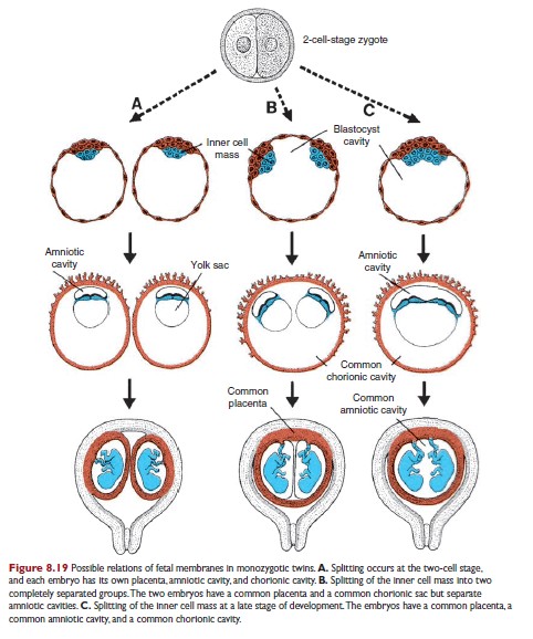
As the name suggests, “Monozygotic” twins develop from a single zygote originating from fertilization of a single oocyte by a single sperm. Hence, these twins are genetically identical. The twinning occurs due to splitting of the zygote at the various stages of the development:
- Separation at 2-celled stage: 2 separate zygotes develop
- Develop like a dizygotic twin (dichorionic and diamniotic) but are genetically and morphologically identical.
- Splitting of embryoblast in early blastocyst stage: 2 separate embryoblast within a single blastocyst cavity
- Have a common placenta and a chorionic cavity but separate amniotic cavities (monochorionic diamniotic).
- Splitting at the bilaminar germ disc stage before the appearance of primitive streak:
- Monochorionic
- Development of 2 primitive streaks within one inner cell mass (trophoblast) – Monoamniotic
Conjoined (Siamese) Twinning
It occurs exactly like monozygotic twins except that there is incomplete “splitting” of primitive streak. Conjoined twins are monoamniotic (i.e., one amnion) and monochorionic (i.e., one chorion).
Concept:
Outer cell mass (Trophoblast) forms chorion and placenta. Inner cell mass (Embryoblast) forms amnion and fetus. If 2 zygotes are implanted separately as in dizygotic twins and monozygotic twins before day 3 – twinning occurs with separate chorion, placenta and amnion. After day 3, there is splitting only of the inner cell mass – hence, the chorion is single but the amions are different. The amniotic cavity begins to be formed at day 8. Hence, splitting after this period leads to twinning with single chorion and single amnion. After 12 days, the partial splitting of primitive streak leads to conjoined twinning.
Dizygotic twins: Dichorionic, Diamniotic
Monozygotic twins:
Day 0-3: Dichorionic, Diamniotic
Day 3-8: Monochorionic, Diamniotic
Day 8-12: Monochorionic, Monoamniotic
>Day 12: Conjoined twins